EARLY CONGENITAL SYPHILIS

Function

Welcome to the Early Congenital Syphilis visual guide. Early congenital syphilis is defined as clinical features that appear during the first 2 years of life. At birth, more than 50% of infants with congenital syphilis are asymptomatic and have no clinical manifestations. With early congenital syphilis there are multiple possible organ systems that can be involved. This visual guide will focus on the following congenital syphilis manifestations: systemic, respiratory, skin, ophthalmologic, abdominal, musculoskeletal, lymphatic, and neurologic.
2 References
  • Cooper JM, Sánchez PJ. Congenital syphilis. Semin Perinatol. 2018;42:176-184.
  • Stafford IA, Workowski KA, Bachmann LH. Syphilis Complicating Pregnancy and Congenital Syphilis. N Engl J Med. 2024;390:242-53.
Healthy Infant

Systemic Manifestations

Healthy Infant

Function

With early congenital syphilis, systemic symptoms are common and include jaundice, fever, edema, and low birth weight. 
Jaundice

Systemic Manifestations

Jaundice

Function

Jaundice caused by hyperbilirubinemia is a common abnormality with early congenital syphilis, and it can result from syphilitic hepatitis and/or hemolysis.
Fever

Systemic Manifestations

Fever

Function

In newborns, temperature is usually taken with a rectal thermometer. A rectal temperature of 100.4 degrees Fahrenheit (38 degrees Celsius) or higher is considered a fever. Fever tends to be more pronounced in infants with congenital syphilis when maternal infection occurs late in pregnancy.
Edema

Systemic Manifestations

Edema

Function

Infants with congenital syphilis who develop nephrotic syndrome typically present at 1–2 months of age with generalized edema (anasarca). In addition, localized edema may occur in association with inflammatory lesions.
Low Birth Weight

Systemic Manifestations

Low Birth Weight

Function

Low birth weight (less than 5 pounds 8 ounces [2,500 grams] is one of the most common features with early congenital syphilis.
Rhinitis

Respiratory Findings

Rhinitis

Function

Rhinitis is a common manifestation of early congenital syphilis that manifests in the first week of life. The rhinitis (nasal discharge) is typically heavy and intractable. The nasal fluid contains abundant spirochetes. This manifestation is also commonly referred to as “snuffles”. The congenital syphilis snuffles manifestation was first described by Michael Underwood and Henry Maunsell in 1789.
 

Details

This photograph shows an infant with early congenital syphilis and purulent rhinitis (“snuffles”).
Figure 3. Infant with Congenital Syphilis and Purulent Rhinitis
This photograph shows an infant with early congenital syphilis and purulent rhinitis (“snuffles”).
Source: Dr. Norman Cole, 1964. Centers for Disease Control and Prevention Public Health Image Library.
Normal Infant Lungs

Respiratory Findings

Normal Infant Lungs

Function

This image shows normal infant lungs without evidence of pneumonia. The inset shows expanded normal right lung.


Pneumonia alba

Respiratory Findings

Pneumonia alba

Function

Pneumonia alba is a process where the newborn alveolar septa become diffusely inflamed and fibrotic in response to Treponema pallidum pulmonary infection. The lungs take on a pale, gray-white appearance. The inset shows an expanded view of pneumonia alba (right lung).
Normal Skin

Skin Findings

Normal Skin

Function

Dermatologic manifestations are very common with early congenital syphilis, occurring in approximately 60-70% of infants with symptomatic congenital syphilis. The congenital syphilis-associated skin abnormalities are often present at birth or very early in life.  
1 Reference
  • Newton J, Silence C, Boetes J, Cohen BA. Mucocutaneous manifestations of congenital syphilis in the neonate: A review of a surging disease. Pediatr Dermatol. 2023;40:238-41.
Diffuse Skin Rash

Skin Findings

Diffuse Skin Rash

Function

A diffuse maculopapular rash is common with congenital syphilis, and it usually appears 1–2 weeks after rhinitis; it is characterized by oval pink lesions that gradually become more reddish-brown or rust-colored. The rash is often most prominent on the chest, back, palms, and soles. The skin lesions contain abundant spirochetes.
 

Details

The rash associated with congenital syphilis often involves the soles and palms (as shown here on the palm of this infant’s right hand). The hand rash often becomes papular, followed by desquamation.  <br />
 
Figure 2. Maculopapular Rash on Hand
The rash associated with congenital syphilis often involves the soles and palms (as shown here on the palm of this infant’s right hand). The hand rash often becomes papular, followed by desquamation.  
 
The congenital syphilis rash can involve the face; if so, it is usually concentrated at the corners of the mouth and nose, with crusting and cracking.<br />
 
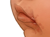
Figure 3. Maculopapular Rash on Face
The congenital syphilis rash can involve the face; if so, it is usually concentrated at the corners of the mouth and nose, with crusting and cracking.
 
Photograph of a 2-month old boy with congenital syphilis showing erythematous macules and papules with some scaling in the upper chest. This bright red rash in this boy represents an early phase of the generalized rash with congenital syphilis; these lesions typically fade to a more copper or rust color.
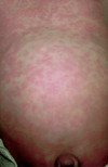
Figure 4. Diffuse Skin Rash
Photograph of a 2-month old boy with congenital syphilis showing erythematous macules and papules with some scaling in the upper chest. This bright red rash in this boy represents an early phase of the generalized rash with congenital syphilis; these lesions typically fade to a more copper or rust color.
Source: Lugo A, Sanchez S, Sanchez JL. Congenital syphilis. Pediatr Dermatol. 2006;23:121-3. Reproduced with permission from Wiley.
Diffuse macular bullous rash on arms, hands, and abdomen of a 1-month old infant with congenital syphilis (Panels A & B).
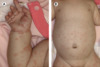
Figure 5. Diffuse Skin Rash in Infant with Congenital Syphilis
Diffuse macular bullous rash on arms, hands, and abdomen of a 1-month old infant with congenital syphilis (Panels A & B).
Source: R Hirate T, Kanda K, Ohshima Y, Shinoda K, Tetsuka N. Congenital syphilis in a 2-month-old infant during Japanese outbreak. Lancet. 2024;404:971. Reproduced with permission from Elsevier.
The congenital syphilis rash often evolves with desquamation. This may involve localized desquamation or diffuse peeling of the skin.
Figure 6. Diffuse Skin Rash with Desquamation on Right Hand
The congenital syphilis rash often evolves with desquamation. This may involve localized desquamation or diffuse peeling of the skin.
This photograph shows a newborn with congenital syphilis shows papulosquamous patches with Biett's collarette of scale on the anterior legs. This description of the collarette of scale was first described by Laurent-Théodore Biett in the early 19<sup>th</sup> century, refers to the narrow rim of peripheral scaling around syphilitic annular maculopapular lesions.
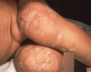
Figure 7. Papulosquamous Patches with Biett's Collarette of Scale in an Infant with Congenital Syphilis
This photograph shows a newborn with congenital syphilis shows papulosquamous patches with Biett's collarette of scale on the anterior legs. This description of the collarette of scale was first described by Laurent-Théodore Biett in the early 19th century, refers to the narrow rim of peripheral scaling around syphilitic annular maculopapular lesions.
Source: Newton J, Silence C, Boetes J, Cohen BA. Mucocutaneous manifestations of congenital syphilis in the neonate: A review of a surging disease. Pediatr Dermatol. 2023;40:238-41. Reproduced with permission from Wiley.
4 References
  • Hirate T, Kanda K, Ohshima Y, Shinoda K, Tetsuka N. Congenital syphilis in a 2-month-old infant during Japanese outbreak. Lancet. 2024;404:971.
  • Kim HY, Kim BJ, Kim JH, Yoo BH. Early congenital syphilis presenting with skin eruption alone: a case report. Korean J Pediatr. 2011;54:512-4.
  • Lugo A, Sanchez S, Sanchez JL. Congenital syphilis. Pediatr Dermatol. 2006;23:121-3.
  • Newton J, Silence C, Boetes J, Cohen BA. Mucocutaneous manifestations of congenital syphilis in the neonate: A review of a surging disease. Pediatr Dermatol. 2023;40:238-41.
Bullous lesions

Skin Findings

Bullous lesions

Function

Bullous lesions are a cutaneous manifestation of congenital syphilis and most often involve the hands and/or feet, but less often they can be more generalized. The bullous lesions, which are referred to as syphilitic pemphigus (or pemphigus syphiliticus), are characterized by large fluid-filled blisters that are fragile and typically rapidly evolve with desquamation and erosions. This manifestation, when present, typically occurs in the first few weeks of life and may even be present at birth.

Details

Extensive bullous rash of soles (image on left) and right palm (image on right) present at birth in an infant with congenital syphilis.
Figure 2. Bullous Palmoplantar Rash on Feet and Hands of Infant with Congenital Syphilis
Extensive bullous rash of soles (image on left) and right palm (image on right) present at birth in an infant with congenital syphilis.
Source: This photograph is reproduced from: Aires Figueiredo L, Botelho Brito T, Labrusco M, Brigham Figueiredo M, Lopo Tuna M. A Rare But Pathognomonic Sign of Congenital Syphilis. J Pediatr. 2023;262:113622. Reproduced with permission from Elsevier.
This photograph shows the plantar surface of the right foot of a neonate with congenital syphilis and pemphigus syphiliticus. There are multiple large blisters in irregular outlines and an erosion on the pad of the fourth toe. 
Figure 3. Bullous Rash on Right Foot of Infant with Congenital Syphilis
This photograph shows the plantar surface of the right foot of a neonate with congenital syphilis and pemphigus syphiliticus. There are multiple large blisters in irregular outlines and an erosion on the pad of the fourth toe. 
Source: This photograph is reproduced from: Alqahtani MH, Alanazi FS, Alqahtani HS, Altowaim AS, Alanzi IA. Early Congenital Syphilis Presenting With Severe Congenital Pneumonia and Cutaneous Manifestations in a Neonate at Birth: A Case Report. Cureus. 2024;16:e69849.
Right foot (Sole) of a neonate with congenital syphilis and multiple blisters in irregular outlines, including some that have ruptured. These features are characteristic of pemphigus syphiliticus.
Figure 4. Bullous Rash with Desquamation on Right Foot of Infant with Congenital Syphilis
Right foot (Sole) of a neonate with congenital syphilis and multiple blisters in irregular outlines, including some that have ruptured. These features are characteristic of pemphigus syphiliticus.
Source: This photograph is reproduced from: Alqahtani MH, Alanazi FS, Alqahtani HS, Altowaim AS, Alanzi IA. Early Congenital Syphilis Presenting With Severe Congenital Pneumonia and Cutaneous Manifestations in a Neonate at Birth: A Case Report. Cureus. 2024;16:e69849.
This photograph shows a large area of desquamation on the dorsal surface of the right foot in a newborn infant with pemphigus syphiliticus.
Figure 5. Pemphigus Syphiliticus with Desquamation on Right Foot of Infant with Congenital Syphilis
This photograph shows a large area of desquamation on the dorsal surface of the right foot in a newborn infant with pemphigus syphiliticus.
Source: Wang EA, Chambers CJ, Silverstein M. A rare presentation of congenital syphilis: Pemphigus syphiliticus in a newborn infant with extensive desquamation of the extremities. Pediatr Dermatol. 2018; 35: e110–e113. Reproduced with permission from Wiley.
This image shows an infant with congenital syphilis and pemphigus syphiliticus. The rash evolved with prominent desquamation of the fingers.
Figure 6. Bullous Rash with Desquamation of Left Hand of Infant with Congenital Syphilis
This image shows an infant with congenital syphilis and pemphigus syphiliticus. The rash evolved with prominent desquamation of the fingers.
Source: This photograph is reproduced from: Alqahtani MH, Alanazi FS, Alqahtani HS, Altowaim AS, Alanzi IA. Early Congenital Syphilis Presenting With Severe Congenital Pneumonia and Cutaneous Manifestations in a Neonate at Birth: A Case Report. Cureus. 2024;16:e69849.
3 References
  • Aires Figueiredo L, Botelho Brito T, Labrusco M, Brigham Figueiredo M, Lopo Tuna M. A Rare But Pathognomonic Sign of Congenital Syphilis. J Pediatr. 2023;262:113622.
  • Alqahtani MH, Alanazi FS, Alqahtani HS, Altowaim AS, Alanzi IA. Early Congenital Syphilis Presenting With Severe Congenital Pneumonia and Cutaneous Manifestations in a Neonate at Birth: A Case Report. Cureus. 2024;16:e69849.
  • Wang EA, Chambers CJ, Silverstein M. A rare presentation of congenital syphilis: Pemphigus syphiliticus in a newborn infant with extensive desquamation of the extremities. Pediatr Dermatol. 2018;35:e110-e113.
Condylomata lata

Skin Findings

Condylomata lata

Function

Condylomata lata are painless, moist, plaque-like, that moist often develop in warm and moist regions of the body (e.g., inner thighs, perianal, axillae, and skin folds). These lesions are typically teeming with spirochetes.
 

Details

This photographs show velvety plaques in the perianal region in a newborn with congenital syphilis.
Figure 3. Condylomata Lata in a Newborn with Congenital Syphilis
This photographs show velvety plaques in the perianal region in a newborn with congenital syphilis.
Source: Petric UB, Ordonez J, Thomas C. Perianal plaques, acral bullae, and desquamation in a neonate. Pediatr Dermatol. 2023;40:718-20. Reproduced with permission from Wiley.
1 Reference
  • Petric UB, Ordonez J, Thomas C. Perianal plaques, acral bullae, and desquamation in a neonate. Pediatr Dermatol. 2023;40:718-20.
Normal Eyes

Ophthalmologic Findings

Normal Eyes

Function

This illustration shows normal infant eyes. There are a number of ophthalmic abnormalities that can develop with syphilis, including cataracts, glaucoma, uveitis, keratitis, iritis, chorioretinitis, and optic neuritis. The following will focus on two common abnormalities: cataracts and uveitis. These abnormalities are often relatively easy to detect on a physical examination.
Cataract

Ophthalmologic Findings

Cataract

Function

Ophthalmologic complications with congenital syphilis can include cataract formation, which appears as an opaque or gray discoloration in the lens of the eye (as shown here in the infant’s right eye). This ocular manifestation would likely cause cloudiness in the infant’s vision. The cataract caused by congenital syphilis can be present at birth or develop early in life.
Uveitis

Ophthalmologic Findings

Uveitis

Function

This illustration shows uveitis, an ophthalmic manifestation of congenital syphilis. Infection in the uvea, which includes the iris, ciliary body, and choroid. Uveitis most often manifests as eye pain and unilateral eye redness (as shown here for the infant’s right eye). There are three types of uveitis depending on the region of the uvea involved: anterior uveitis (iritis), intermediate uveitis, posterior, or panuveitis.
Cataract and Uveitis

Ophthalmologic Findings

Cataract and Uveitis

Function

Some infants with congenital syphilis may have multiple ocular abnormalities, as shown here with both cataract (white opaque area on lens of the right eye) and uveitis (diffuse redness in the right eye). If both of these manifestations are present, the infant would likely have a significant decrease in vision.
Normal Liver and Spleen

Abdominal Findings

Normal Liver and Spleen

Function

This image shows the typical appearance and size of the liver and spleen in a healthy newborn.
Hepatomegaly

Abdominal Findings

Hepatomegaly

Function

Hepatomegaly is a common abnormality with congenital syphilis. This is often detected on fetal ultrasound. Hepatomegaly may occur with or without splenomegaly. In this image, the infant does not have splenomegaly.
Hepatosplenomegaly

Abdominal Findings

Hepatosplenomegaly

Function

Among infants with congenital syphilis who have hepatomegaly, approximately 50% also have splenomegaly. Isolated splenomegaly typically does not occur.
Normal Musculoskeletal

Musculoskeletal Findings

Normal Musculoskeletal

Function

Abnormalities of the long bones occur in 60-80% of cases of untreated congenital syphilis. The bone manifestations are usually bilateral and include metaphyseal changes, bone destruction, periosteal inflammation, and pseudoparalysis.
1 Reference
  • Rasool MN, Govender S. The skeletal manifestations of congenital syphilis. A review of 197 cases. J Bone Joint Surg Br. 1989;71:752-5.
Inflammation and Bone Destruction

Musculoskeletal Findings

Inflammation and Bone Destruction

Function

The illustration shows inflammation of the distal metaphysis of the femur and destruction of the medial metaphysis of the proximal left tibia. Bilateral proximal medial metaphyseal tibial bone destruction with congenital syphilis was originally described by Hans Wimberger in 1925 and this finding is referred to as Wimberger’s sign.
 

Details

This inset shows a close-up view of inflammation of the distal metaphysis of the femur (top black arrow) and destruction of the medial metaphysis of the proximal left tibia (bottom black arrow).<br />
<br />
 
Figure 2. Osteochondritis and Bone Destruction (Inset)
This inset shows a close-up view of inflammation of the distal metaphysis of the femur (top black arrow) and destruction of the medial metaphysis of the proximal left tibia (bottom black arrow).

 
This radiograph, from an infant with congenital syphilis, shows multiple bony changes, including destruction of the medial metaphysis of the left tibia (top white arrow) and diffuse tibial periosteal reaction (bottom white arrow). These findings illustrate Wimberger sign
Figure 3. Radiograph with Wimberger's Sign
This radiograph, from an infant with congenital syphilis, shows multiple bony changes, including destruction of the medial metaphysis of the left tibia (top white arrow) and diffuse tibial periosteal reaction (bottom white arrow). These findings illustrate Wimberger sign
Source: reprinted from Stephens JR and Arenth, J.  Wimberger Sign in Congenital Syphilis. J Pediatr. 2015;167:1451. Copyright 2015 (reproduced with permission from Elsevier).
2 References
  • Mannelli L, Perez FA, Parisi MT, Giacani L. A case of congenital syphilis. Emerg Radiol. 2013;20:337-9.
  • Stephens JR, Arenth J. Wimberger Sign in Congenital Syphilis. J Pediatr. 2015;167:1451.
Periosteal Inflammation

Musculoskeletal Findings

Periosteal Inflammation

Function

The illustration shows periosteal inflammation of the proximal left tibia (arrow). These lesions may be associated with significant pain. Periostitis usually involves multiple regions of the metaphyses of long bones (inset) and abnormal long bone radiographic findings are nearly always present.
 
Pseudoparalysis of Parrot

Musculoskeletal Findings

Pseudoparalysis of Parrot

Function

Infants with congenital syphilis and periostitis may experience pain and limited movement of the affected limbs. If the periostitis is severe, the infant may entirely avoid moving the affected limb and this condition can be mistaken for paralysis, hence the term "pseudoparalysis". This was originally described by Joseph Marie Jules Parrot in 1871 and the condition is commonly referred to as Pseudoparalysis of Parrot. Typically, the upper limbs are affected, with bilateral involvement. The infant clinically presents with the upper extremities hanging flaccid and extended along the side of the body.
 

Details

2 References
  • Kocher MS, Caniza M. Parrot pseudoparalysis of the upper extremities. A case report. J Bone Joint Surg Am. 1996;78:284-7.
  • Pereira AA, Castro SM, Venturini RR, César FO, Fortes PM, Costa PS. Pseudoparalysis of Parrot: A Diagnostic Aid in Congenital Syphilis. J Pediatr. 2017;190:282.
Lymphadenopathy

Lymph Node Findings

Lymphadenopathy

Function

Non-tender lymphadenopathy is a common manifestation of early congenital syphilis. Lymphadenopathy associated with congenital syphilis is usually generalized and nontender. The lymphadenopathy develops in response to Treponema pallidum infection in the lymph nodes. The most common areas of involvement are the neck, axillae, and groin.
 
Neurologic Findings

Neurologic Findings

Neurologic Findings

Function

Central nervous system infection with Treponema pallidum is common among infants with confirmed congenital syphilis. There are an array of neurologic manifestations that can occur with early congenital syphilis, including syphilitic leptomeningitis, meningoencephalitis, cranial nerve palsies, seizures, sensorineural hearing loss, and hydrocephalus.
1 Reference
  • Michelow IC, Wendel GD Jr, Norgard MV, et al. Central nervous system infection in congenital syphilis. N Engl J Med. 2002;346:1792-8.

Share by e-mail

Page Not Found

Unfortunately the page you requested could not be found on this server. Please try again.