Disclaimer: The NSTDC Image Library and Featured Quiz contain photographs and illustrations that may be graphic in nature. These are intended for educational purposes only.
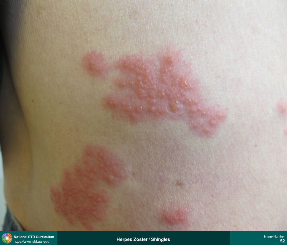
Herpes Zoster / Shingles
Herpes zoster (shingles) in the lower back region.
Photo: Erythema, Vesicle / Vesicles, Back, Light skin tone, Painful
Courtesy of Negusse Ocbamichael, PA
Erythema, Vesicle / Vesicles Back, Light skin tone
52
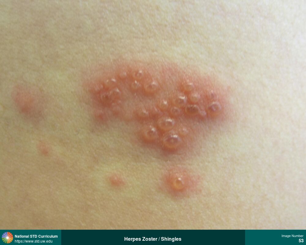
Herpes Zoster / Shingles
Photo: Erythema, Vesicle / Vesicles, Back, Light skin tone, Painful
Courtesy of Negusse Ocbamichael, PA
Erythema, Vesicle / Vesicles Back, Light skin tone
53
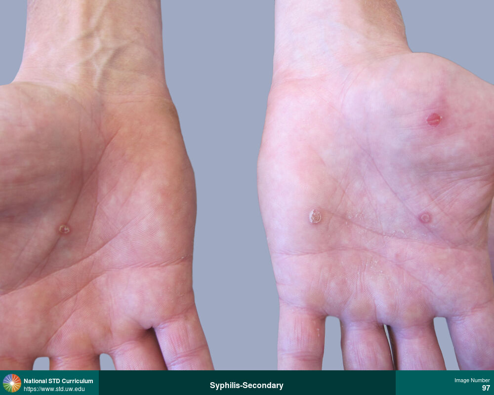
Syphilis-Secondary
Papular lesions with collarette of scale on the palms of both hands caused by secondary syphilis.
Photo: Annular, Hyperpigmentation, Papule / Papules, Hand (Left), Hand (Right), Hands, Non-Painful
Courtesy of Negusse Ocbamichael, PA
Annular, Hyperpigmentation, Papule / Papules Hand (Left), Hand (Right), Hands
97
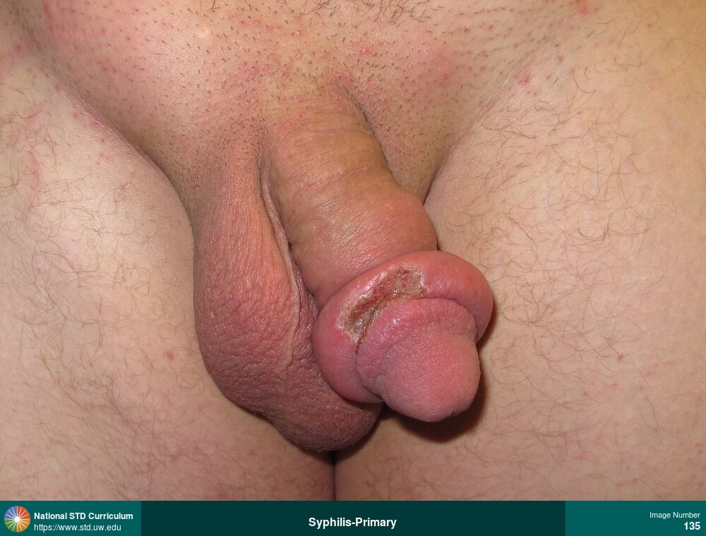
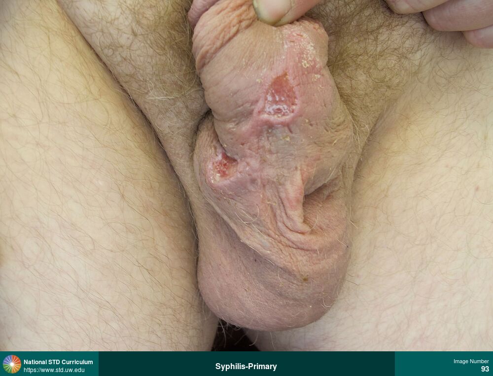
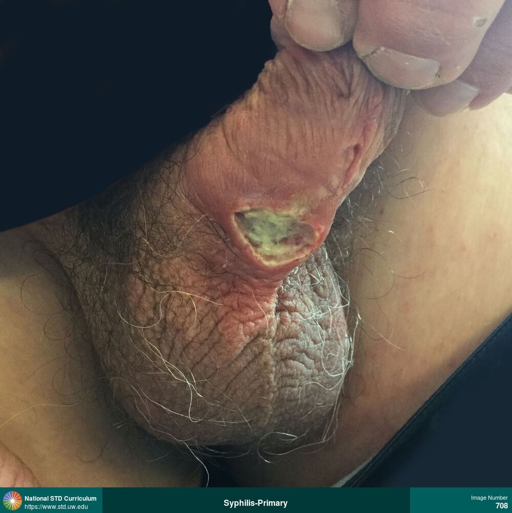
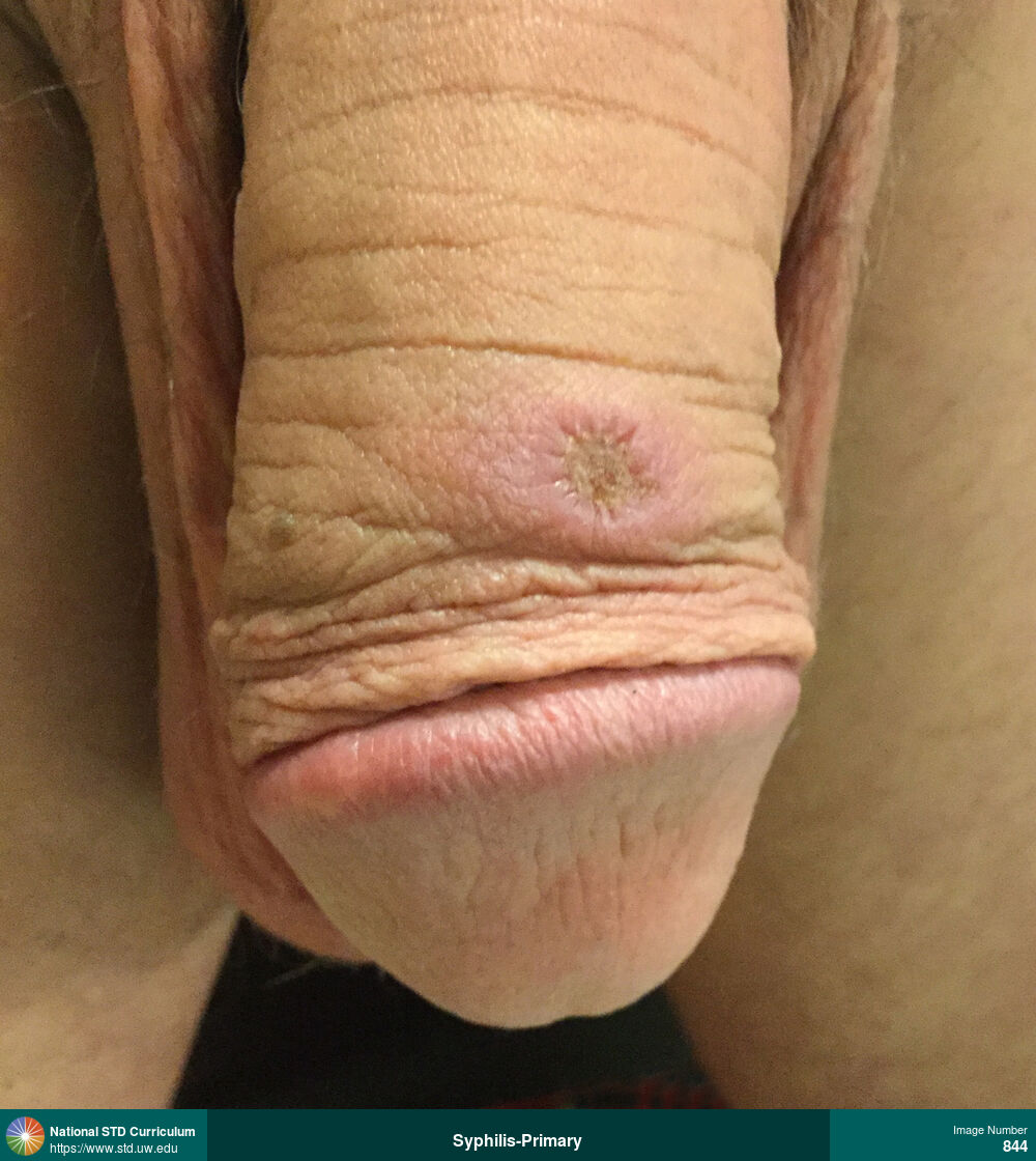
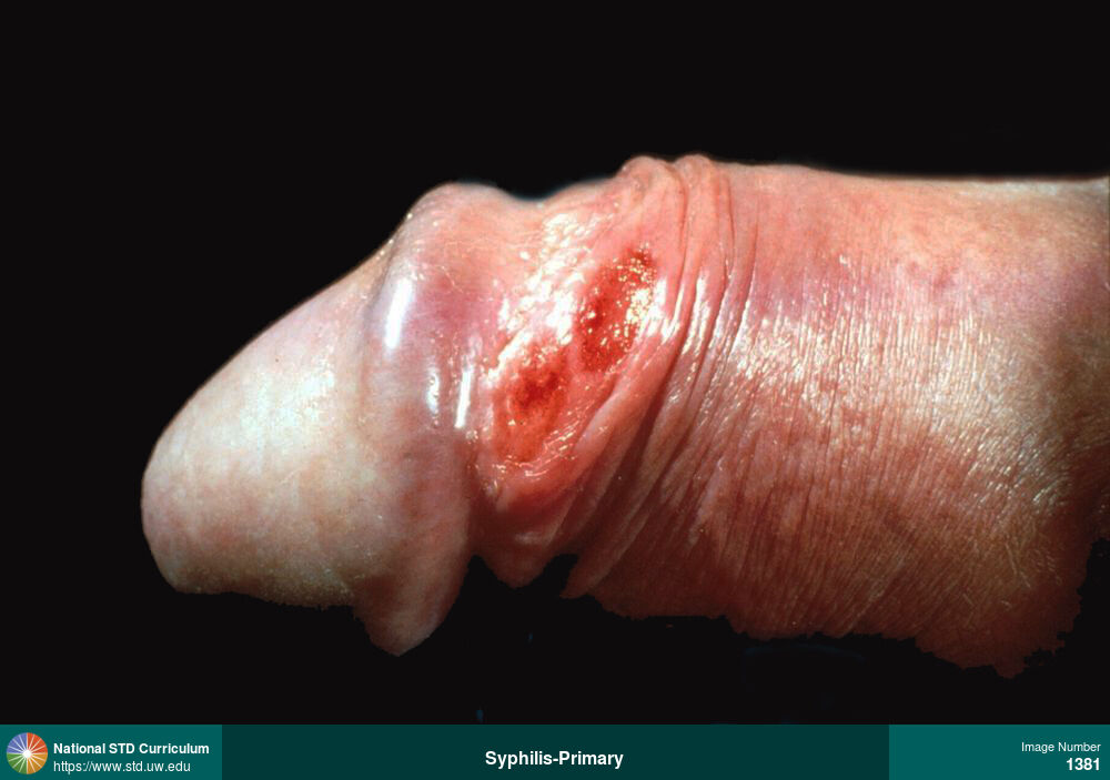
Syphilis-Primary, Unknown (Syphilis)
Large primary syphilis ulcer on distal shaft of penis with marked edema.
Photo: Adenopathy, Edema / Swelling, Ulcer / Ulcers, Light skin tone, Penis
Courtesy of Negusse Ocbamichael, PA
Adenopathy, Edema / Swelling, Ulcer / Ulcers Light skin tone, Penis
135
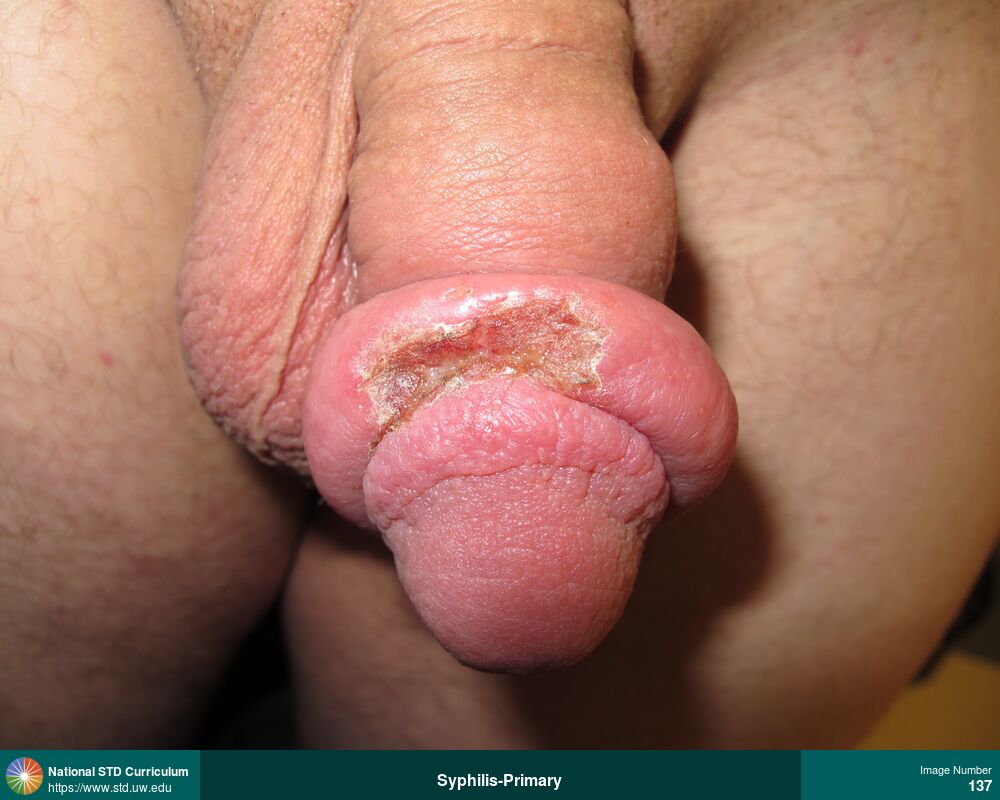
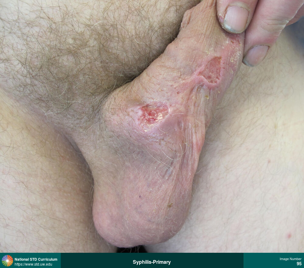
Syphilis-Primary, Unknown (Syphilis)
Large primary syphilis ulcer on distal shaft of penis with marked edema.
Photo: Edema / Swelling, Ulcer / Ulcers, Light skin tone, Penis
Courtesy of Negusse Ocbamichael, PA
Edema / Swelling, Ulcer / Ulcers Light skin tone, Penis
137
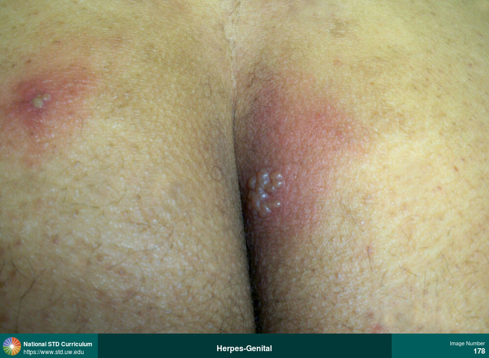
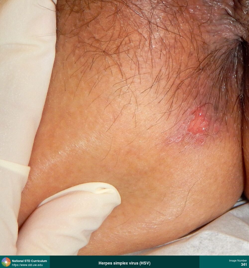
Herpes-Genital
Clusters of vesicles with surrounding erythema on the left buttocks and gluteal fold region caused by recurrent herpes simplex virus (HSV) infection.
Photo: Edema / Swelling, Erythema, Vesicle / Vesicles, Buttock, Light skin tone, Painful, Rash
Courtesy of Christine M. Johnston, MD, MPH
Edema / Swelling, Erythema, Vesicle / Vesicles Buttock, Light skin tone
178
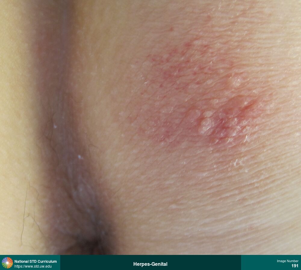
Herpes-Genital
Cluster of vesicular lesions with surrounding erythema in perianal/gluteal region caused by herpes simplex virus (HSV) infection.
Photo: Erythema, Vesicle / Vesicles, Buttock, Light skin tone, Painful, Rash
Courtesy of Christine M. Johnston, MD, MPH
Erythema, Vesicle / Vesicles Buttock, Light skin tone
191
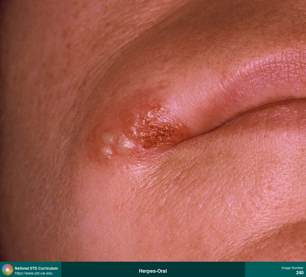
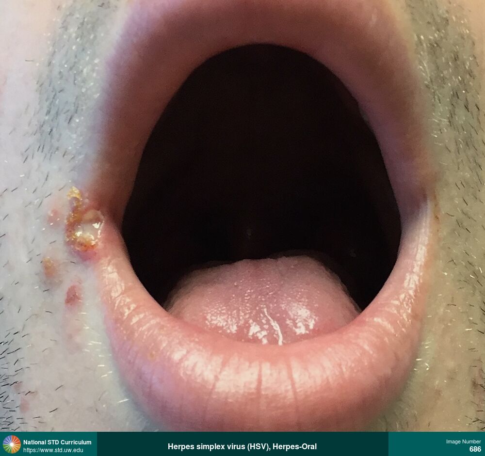
Herpes-Oral
Oral HSV
Photo: Erythema, Ulcer / Ulcers, Vesicle / Vesicles, Head, Lips, Painful
Courtesy of Christine M. Johnston, MD, MPH
Erythema, Ulcer / Ulcers, Vesicle / Vesicles Head, Lips
240
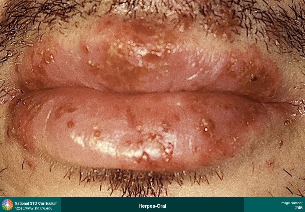
Herpes-Oral
Extensive vesicular lesions involving all regions of lips in a man with first episode (primary) oral herpes simplex virus (HSV) infection.
Photo: Erythema, Ulcer / Ulcers, Vesicle / Vesicles, Lips, Painful
Courtesy of Christine M. Johnston, MD, MPH
Erythema, Ulcer / Ulcers, Vesicle / Vesicles Lips
245
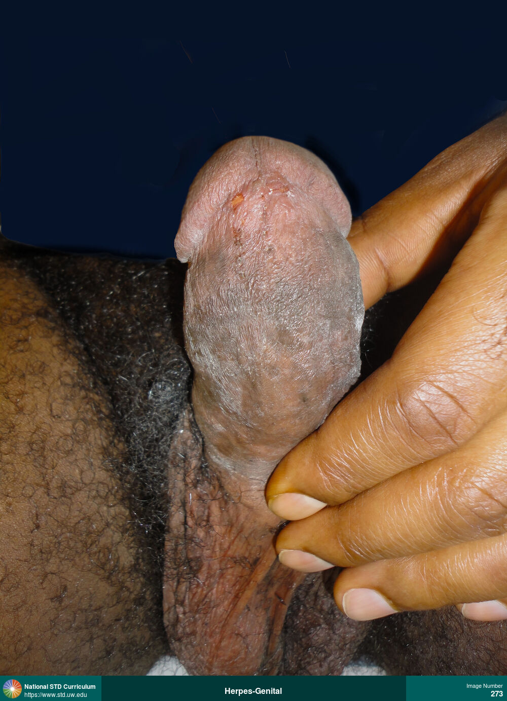
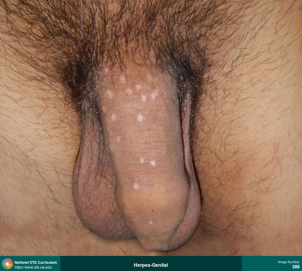
Herpes-Genital
Multiple hypopigmented healing genital herpes lesions on the shaft of the penis.
Photo: Hypopigmentation, Macule / Macules, Light skin tone, Penis
Courtesy of Christine M. Johnston, MD, MPH
Hypopigmentation, Macule / Macules Light skin tone, Penis
288
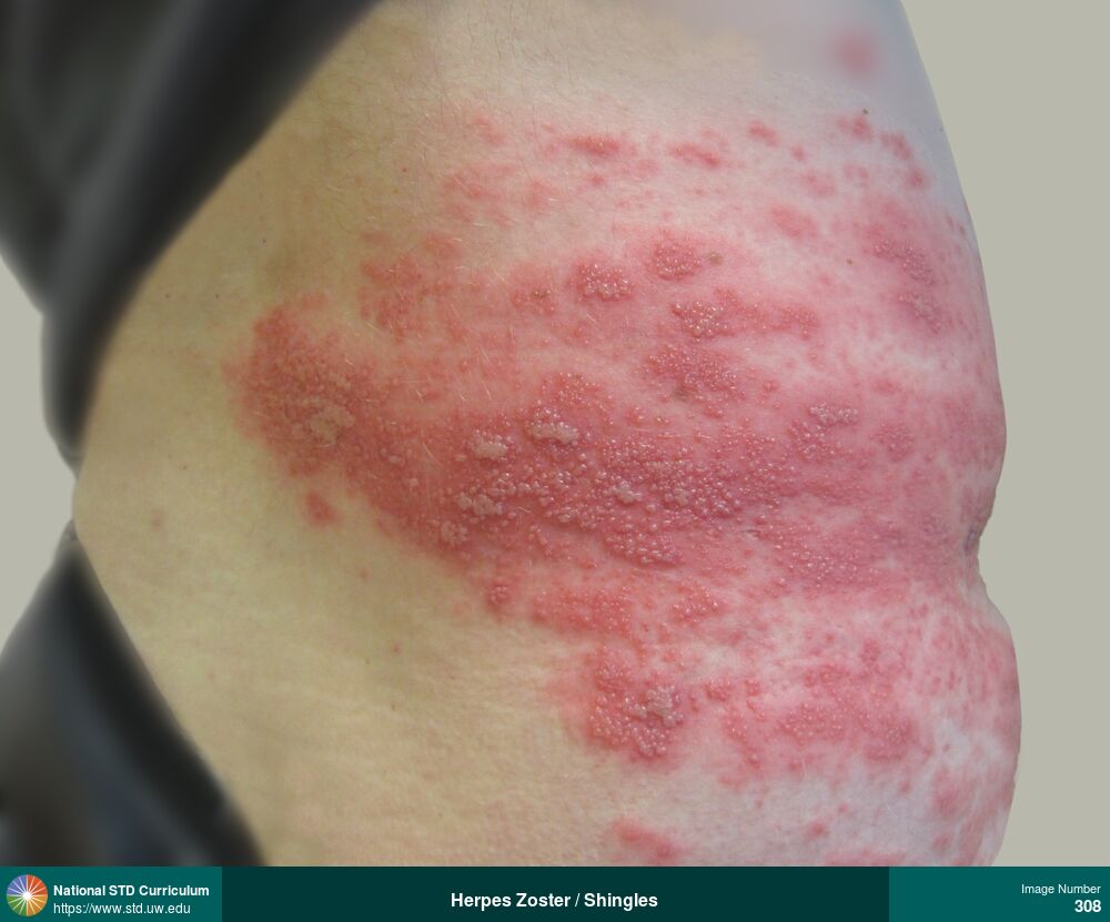
Herpes Zoster / Shingles, Zoster / Shingles
Dense, extensive clusters of vesicles with surrounding erythema on right flank and abdominal region caused by herpes zoster (shingles).
Photo: Erythema, Vesicle / Vesicles, Abdomen, Back, Painful
Courtesy of Christine M. Johnston, MD, MPH
Erythema, Vesicle / Vesicles Abdomen, Back
308
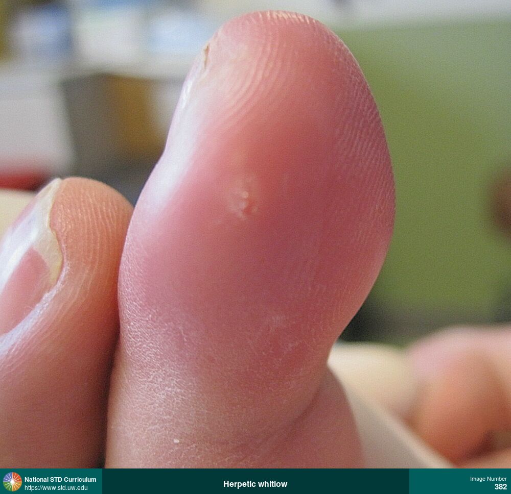
Herpes-Digital (Whitlow)
Herpetic whitlow
Photo: Edema / Swelling, Erythema, Whitlow, Hand (Right), Light skin tone, Painful
Courtesy of Christine M. Johnston, MD, MPH
Edema / Swelling, Erythema, Whitlow Hand (Right), Light skin tone
382

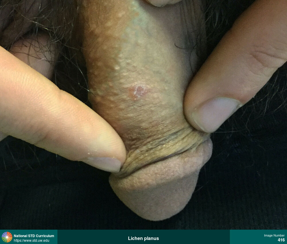
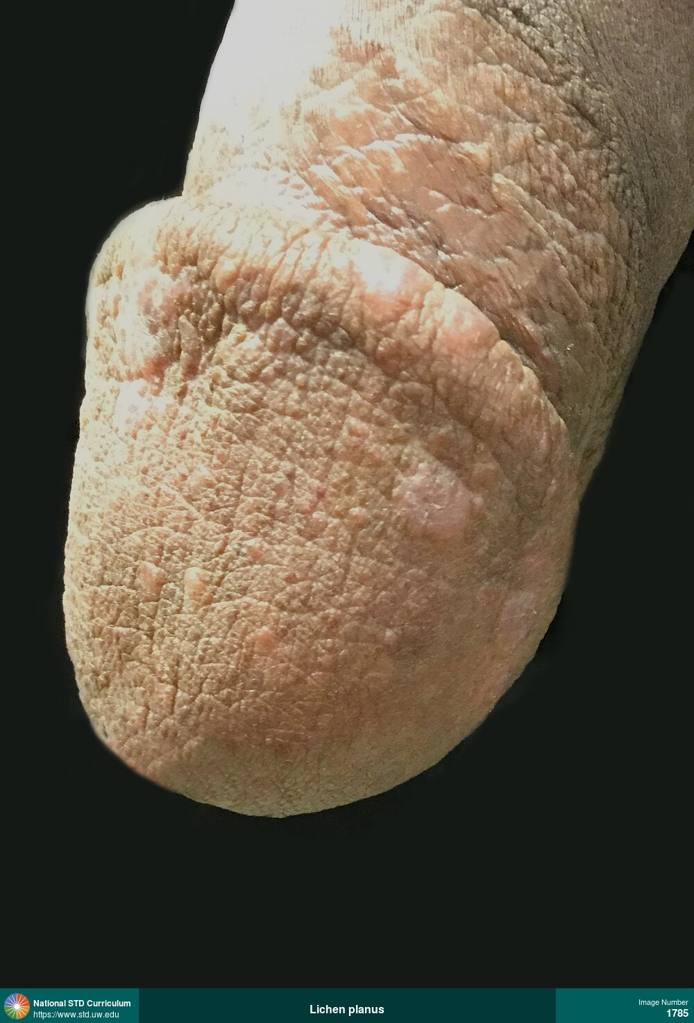
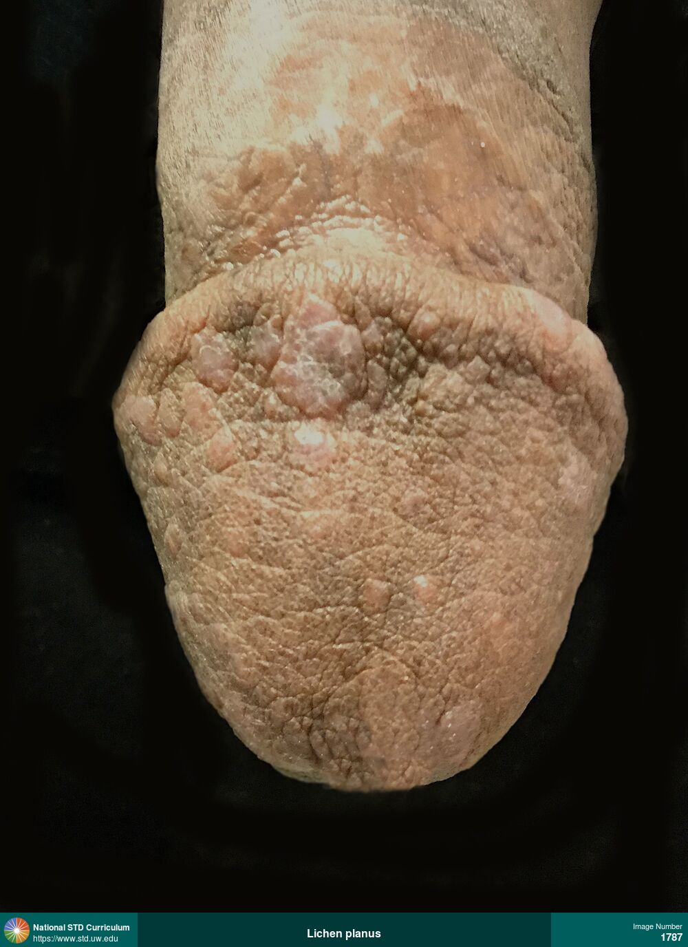
Lichen planus
Annular lesion with white raised, lacy/reticulated border (often referred to as Wickam's striae) and darker center on the glans penis caused by lichen planus.
Photo: Annular, Patch/Patches, Plaque, Penis, Non-Painful
Courtesy of Negusse Ocbamichael, PA
Annular, Patch/Patches, Plaque Penis
413
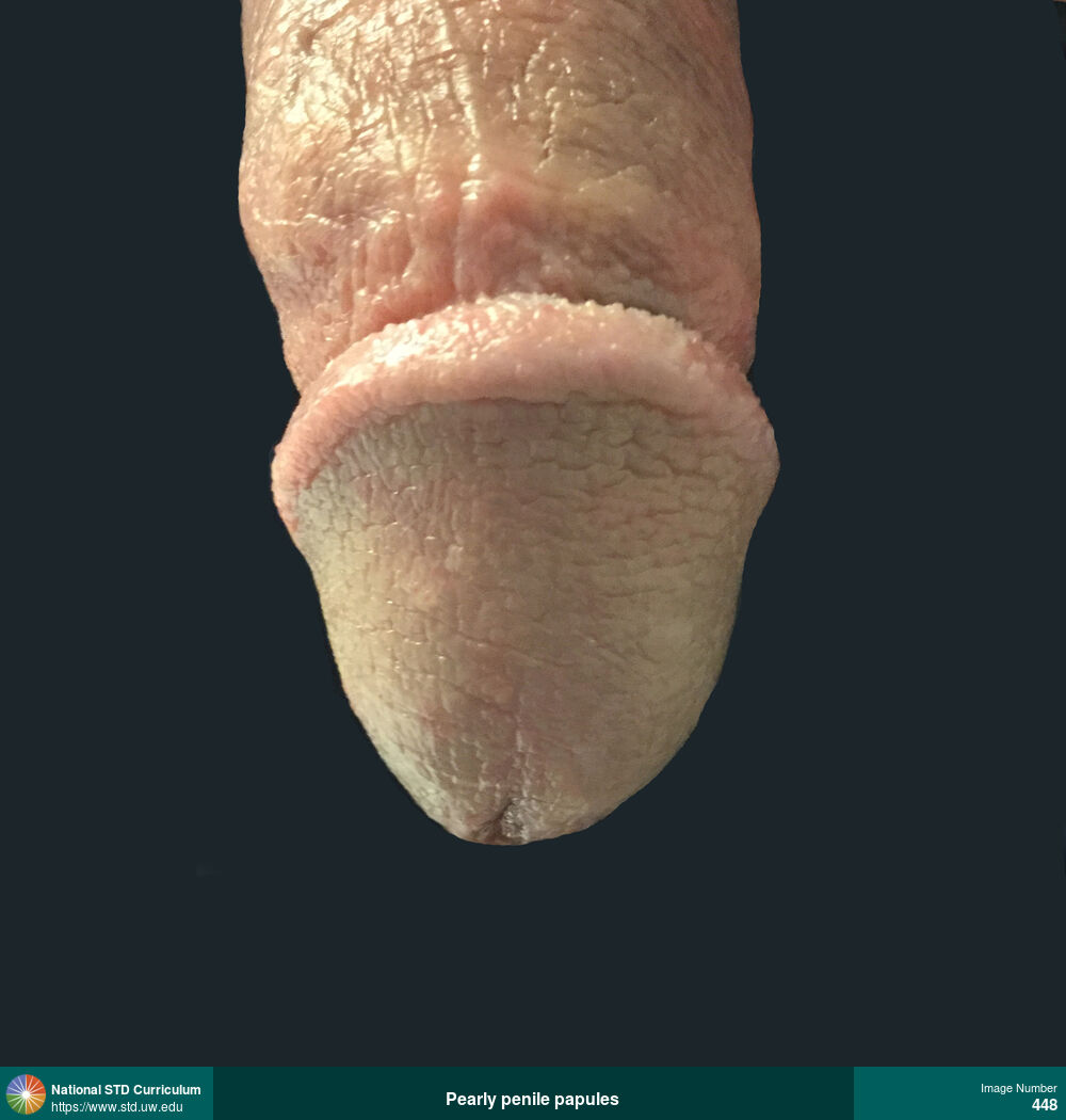
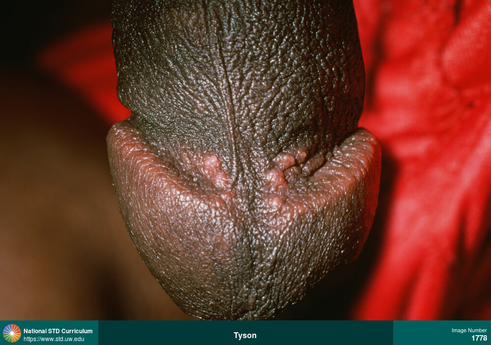
Balanitis, Pearly penile papules
Pearly penile papules are a normal anatomic variant that appear as small (<1 mm), flesh-colored bumps in rows around the corona of the penis.
Photo: Papule / Papules, Light skin tone, Penis, Non-Painful
Courtesy of Negusse Ocbamichael, PA
Papule / Papules Light skin tone, Penis
448
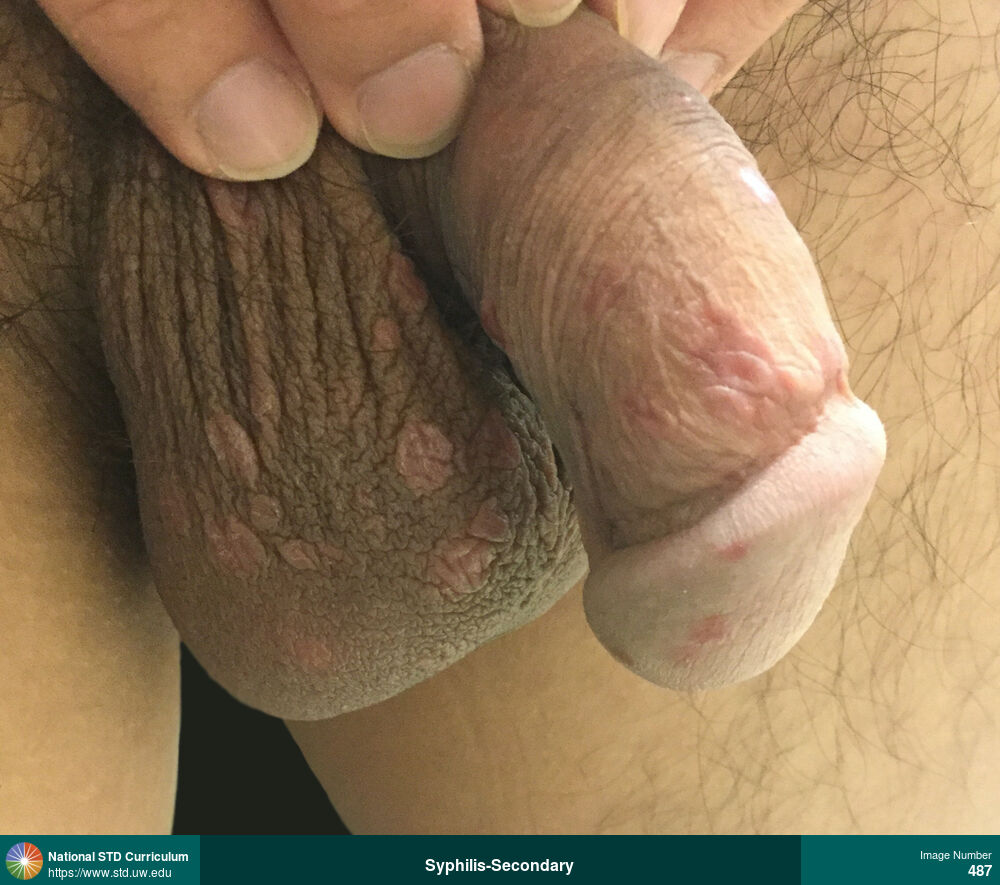
Syphilis-Secondary
Multiple raised papular lesions on penis and scrotum consistent with condylomata lata in a man with secondary syphilis.
Photo: Macule / Macules, Papule / Papules, Rash, Light skin tone, Penis, Scrotum, Rash
Courtesy of Negusse Ocbamichael, PA
Macule / Macules, Papule / Papules, Rash Light skin tone, Penis, Scrotum
487
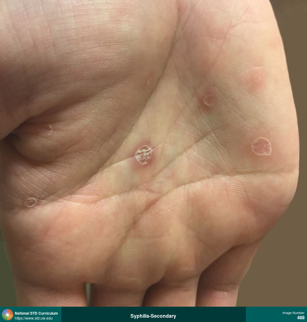
Syphilis-Secondary
Papular rash with scale on the palm of the right hand caused by secondary syphilis.
Photo: Macule / Macules, Papule / Papules, Plaque, Rash, Scale, Hand (Left), Hand (Right), Light skin tone
Courtesy of Negusse Ocbamichael, PA
Macule / Macules, Papule / Papules, Plaque, Rash, Scale Hand (Left), Hand (Right), Light skin tone
488
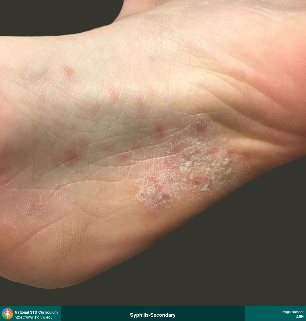
Syphilis-Secondary
Papular rash with scale on the sole of the right foot caused by secondary syphilis.
Photo: Macule / Macules, Papule / Papules, Plaque, Rash, Scale, Feet/Soles, Light skin tone
Courtesy of Negusse Ocbamichael, PA
Macule / Macules, Papule / Papules, Plaque, Rash, Scale Feet/Soles, Light skin tone
489
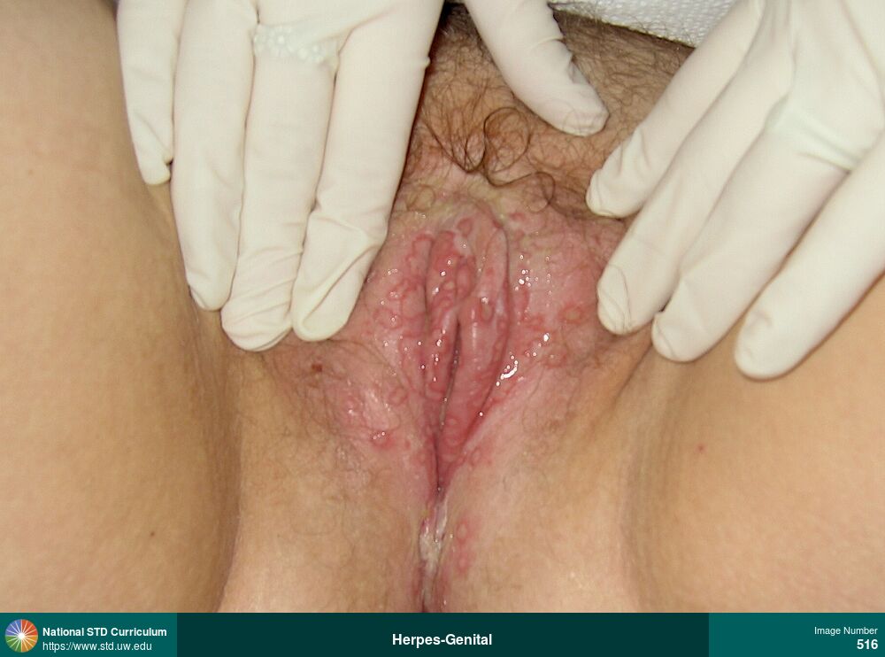
Herpes-Genital
Multiple extensive vulvar and labial ulcerations (with surrounding ring of erythema) caused by first-episode (primary) herpes simplex virus (HSV) infection.
Photo: Erythema, Lesion, Ulcer / Ulcers, Labia (majora/minora), Light skin tone, Vulva, Painful
Courtesy of Christine M. Johnston, MD, MPH
Erythema, Lesion, Ulcer / Ulcers Labia (majora/minora), Light skin tone, Vulva
516
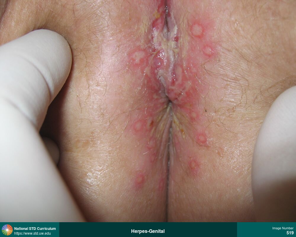
Herpes-Genital
Multiple perianal ulcerated lesions with surrounding ring of erythema caused by herpes simplex virus (HSV) infection.
Photo: Lesion, Ulcer / Ulcers, Anal / Perianal, Light skin tone, Vulva, Painful
Courtesy of Christine M. Johnston, MD, MPH
Lesion, Ulcer / Ulcers Anal / Perianal, Light skin tone, Vulva
519
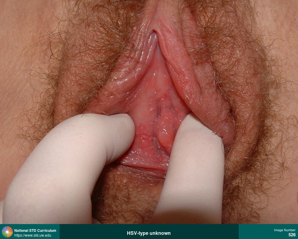
Herpes-Genital
Photo: Lesion, Ulcer / Ulcers, Light skin tone, Vulva, Painful
Courtesy of Christine M. Johnston, MD, MPH
Lesion, Ulcer / Ulcers Light skin tone, Vulva
526

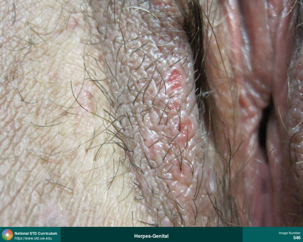
Herpes-Genital
Solitary ulcer on lower left labia majora caused by recurrent herpes simplex virus (HSV) infection.
Photo: Lesion, Ulcer / Ulcers, Dark skin tone, Labia (majora/minora), Painful
Courtesy of Christine M. Johnston, MD, MPH
Lesion, Ulcer / Ulcers Dark skin tone, Labia (majora/minora)
532
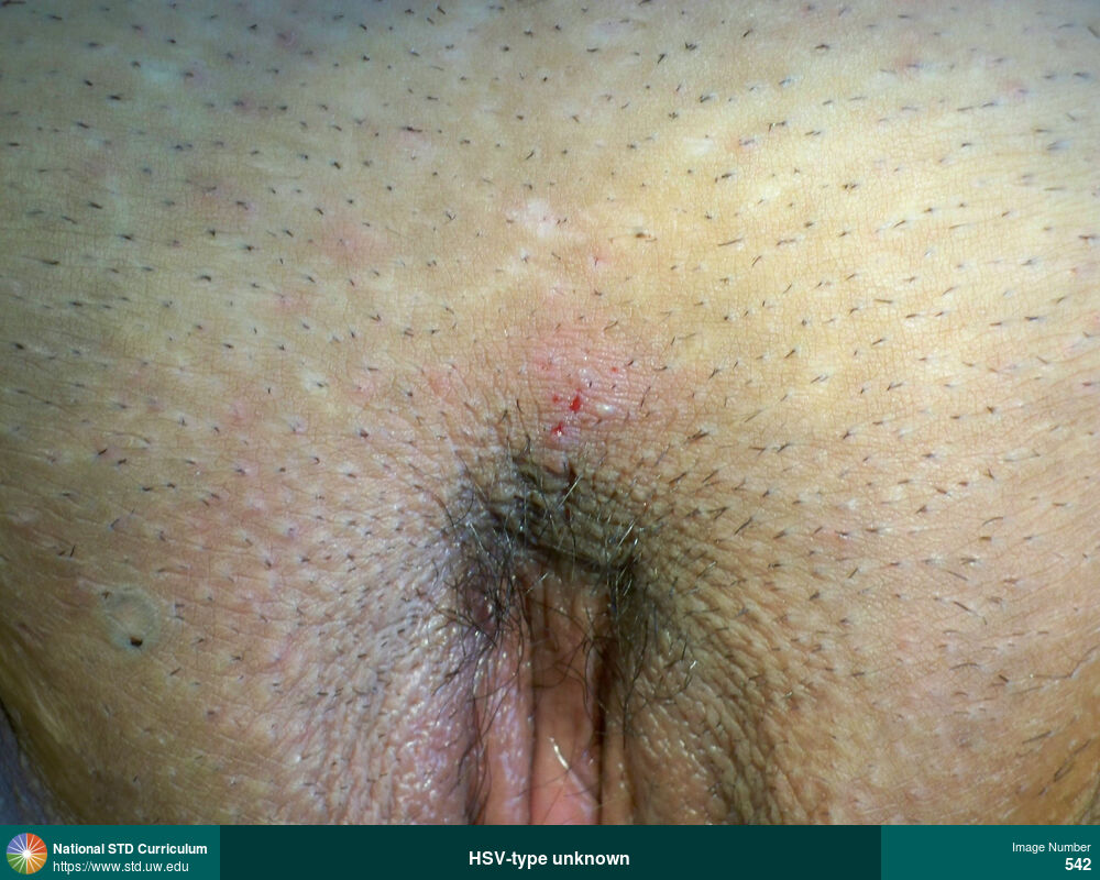
Herpes-Genital
Cluster of vesicles and ulcerated lesions on vulva caused by herpes simplex virus (HSV) infection
Photo: Lesion, Ulcer / Ulcers, Vesicle / Vesicles, Light skin tone, Vulva
Courtesy of Christine M. Johnston, MD, MPH
Lesion, Ulcer / Ulcers, Vesicle / Vesicles Light skin tone, Vulva
542
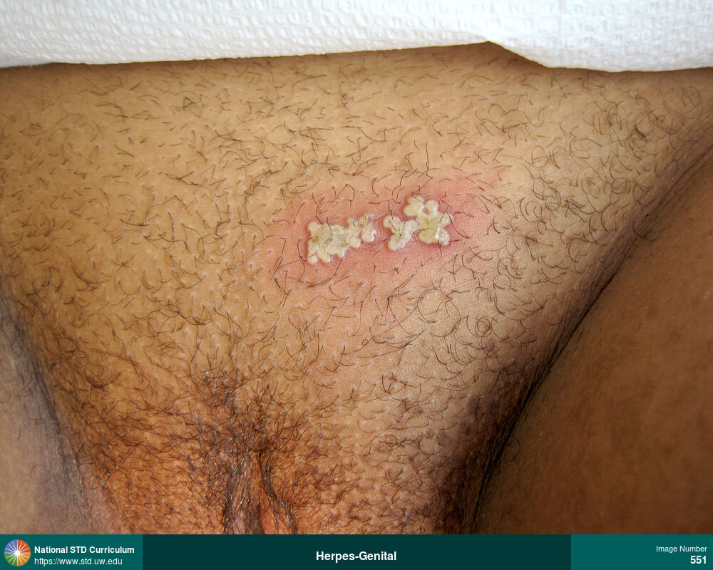
Herpes-Genital
Cluster of vesicles with surrounding erythema on suprapubic region caused by recurrent herpes simplex virus (HSV) infection.
Photo: Erythema, Papule / Papules, Vesicle / Vesicles, Abdomen, Dark skin tone, Suprapubic (Hypogastrium), Painful
Courtesy of Christine M. Johnston, MD, MPH
Erythema, Papule / Papules, Vesicle / Vesicles Abdomen, Dark skin tone, Suprapubic (Hypogastrium)
551

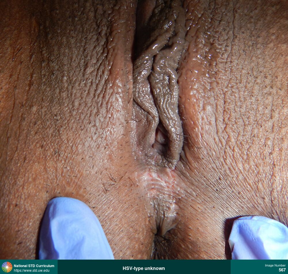
Herpes-Genital
Linear superficial ulceration in lower vulvar/perianal region caused by recurrent herpes simplex virus (HSV) infection.
Photo: Lesion, Ulcer / Ulcers, Anal / Perianal, Dark skin tone, Vulva
Courtesy of Christine M. Johnston, MD, MPH
Lesion, Ulcer / Ulcers Anal / Perianal, Dark skin tone, Vulva
567
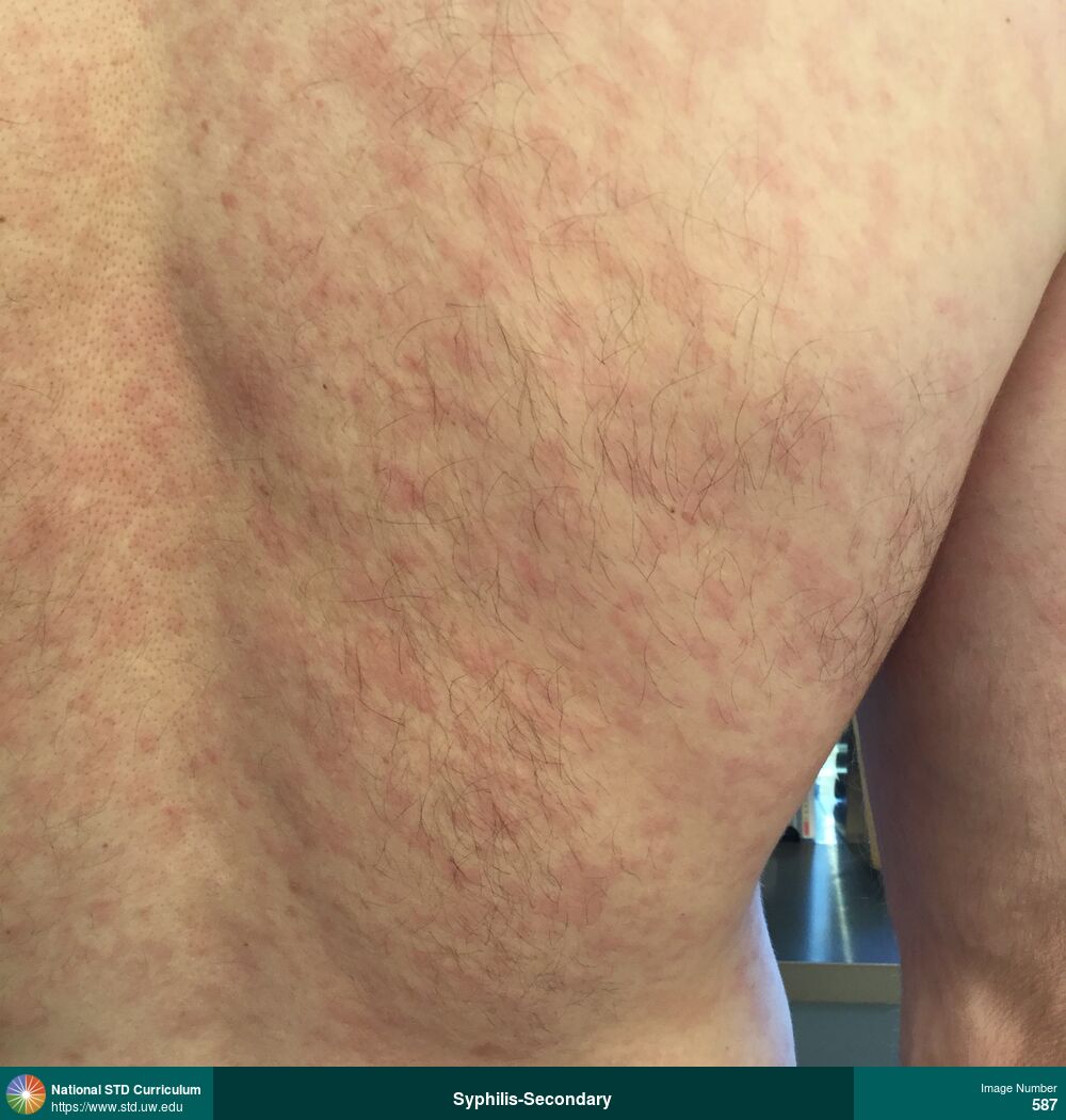
Syphilis-Secondary
Diffuse, erythematous macular rash on back caused by secondary syphilis.
Photo: Erythema, Macule / Macules, Rash, Back, Light skin tone, Itch, Non-Painful, Rash
Courtesy of Negusse Ocbamichael, PA
Erythema, Macule / Macules, Rash Back, Light skin tone
587

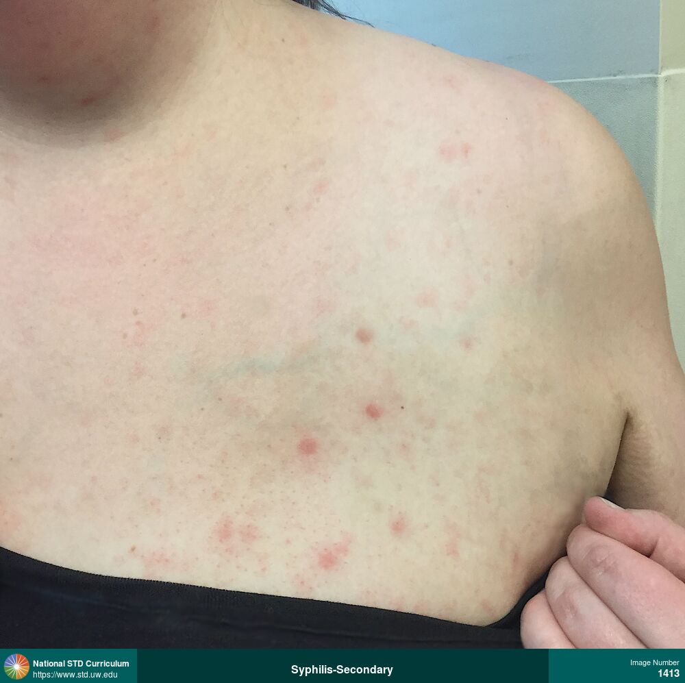
Syphilis-Secondary
Diffuse, erythematous macular rash on anterior chest and abdomen caused by secondary syphilis.
Photo: Erythema, Macule / Macules, Abdomen, Chest, Light skin tone, Itch, Non-Painful, Rash
Courtesy of Negusse Ocbamichael, PA
Erythema, Macule / Macules Abdomen, Chest, Light skin tone
589
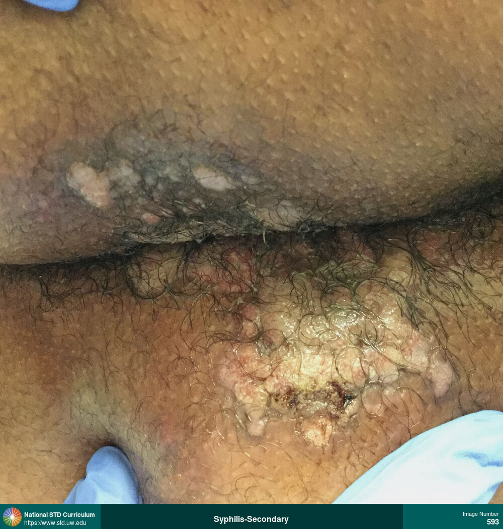
HPV-Related Cancers, Syphilis-Secondary
Large, moist, verrucous, plaque-like lesions in gluteal folds cause by secondary syphilis (condylomata lata).
Photo: Plaque, Verrucous, Anal / Perianal, Buttock, Dark skin tone
Courtesy of Negusse Ocbamichael, PA
Plaque, Verrucous Anal / Perianal, Buttock, Dark skin tone
593
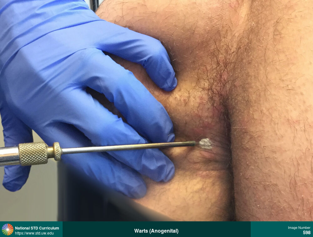
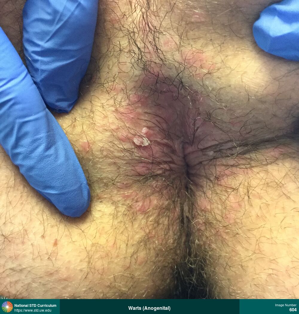
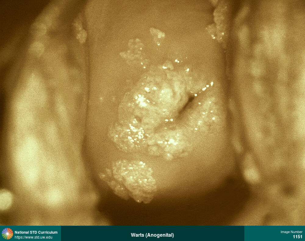
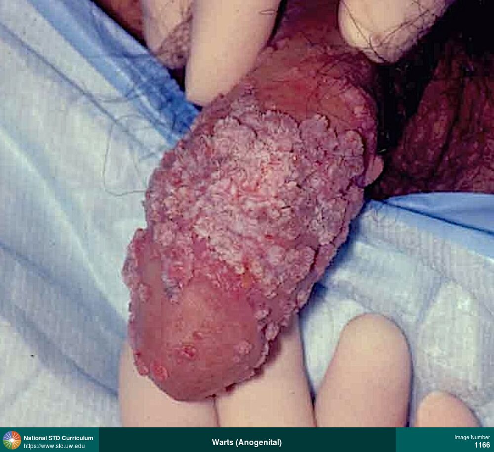
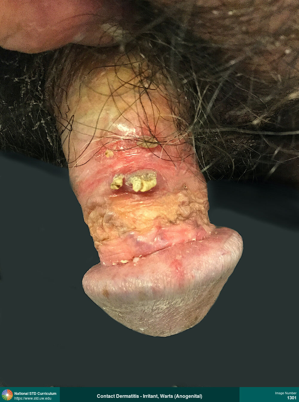
HPV-Related Cancers, Warts (Anogenital)
Perianal warts.
Photo: Verrucous, Anal / Perianal, Buttock
Courtesy of Negusse Ocbamichael, PA
Verrucous Anal / Perianal, Buttock
604
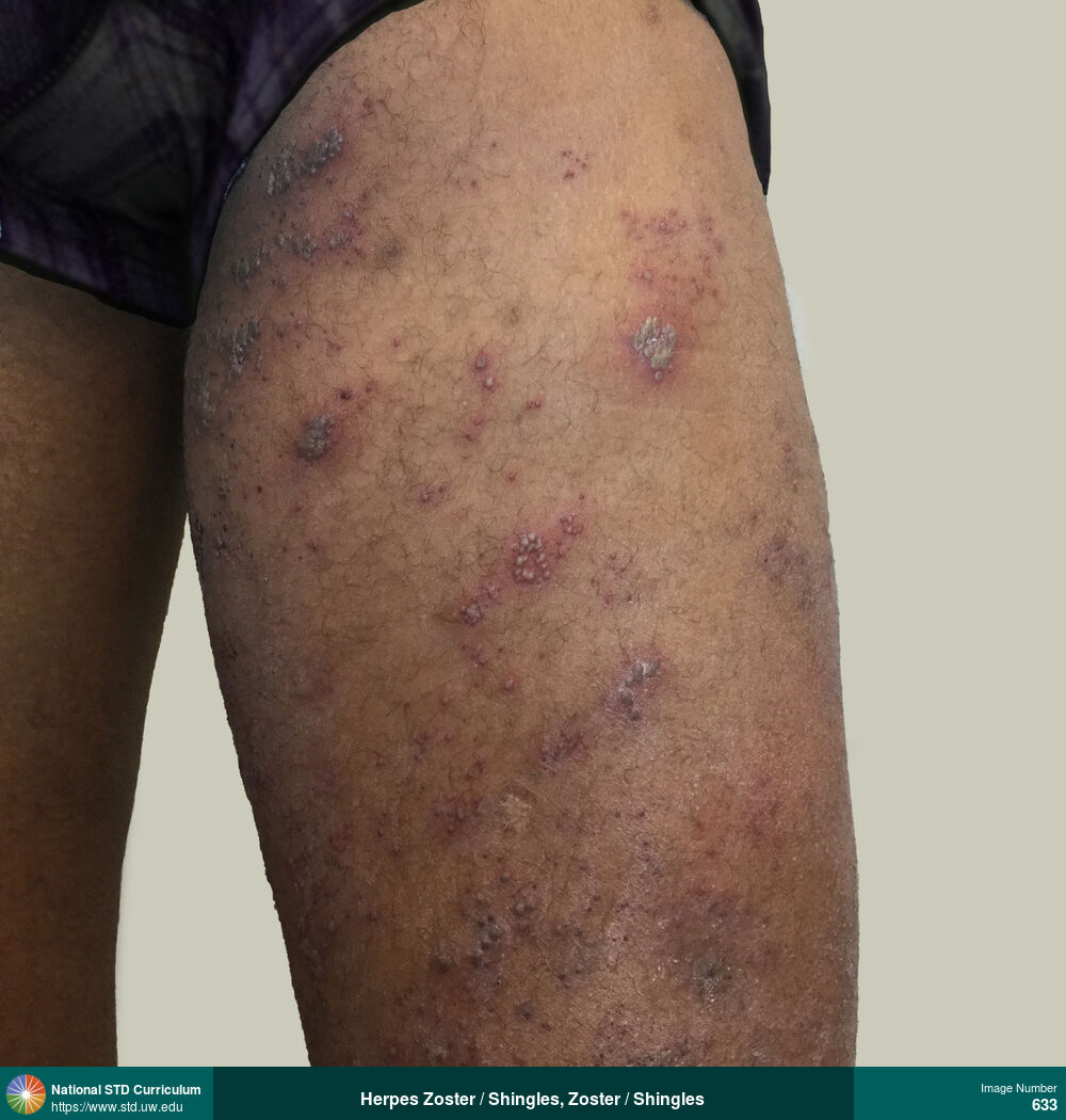
Herpes Zoster / Shingles, Zoster / Shingles
Painful vesicular rash on left anterior thigh caused by herpes zoster (shingles). The rash occurred in the distribution of the anterior femoral cutaneous nerve (L2 and L3).
Photo: Erythema, Vesicle / Vesicles, Dark skin tone, Thigh
Courtesy of Negusse Ocbamichael, PA
Erythema, Vesicle / Vesicles Dark skin tone, Thigh
633

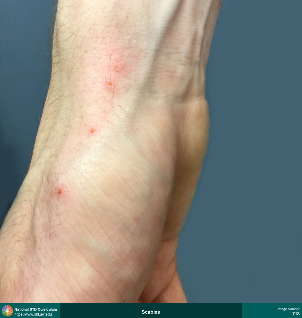
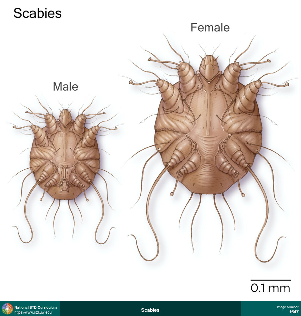
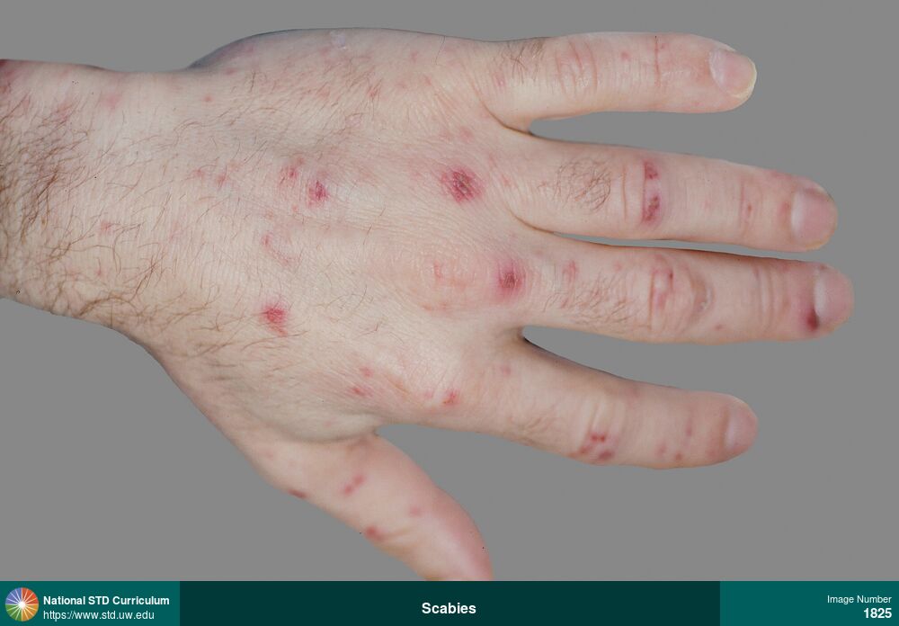
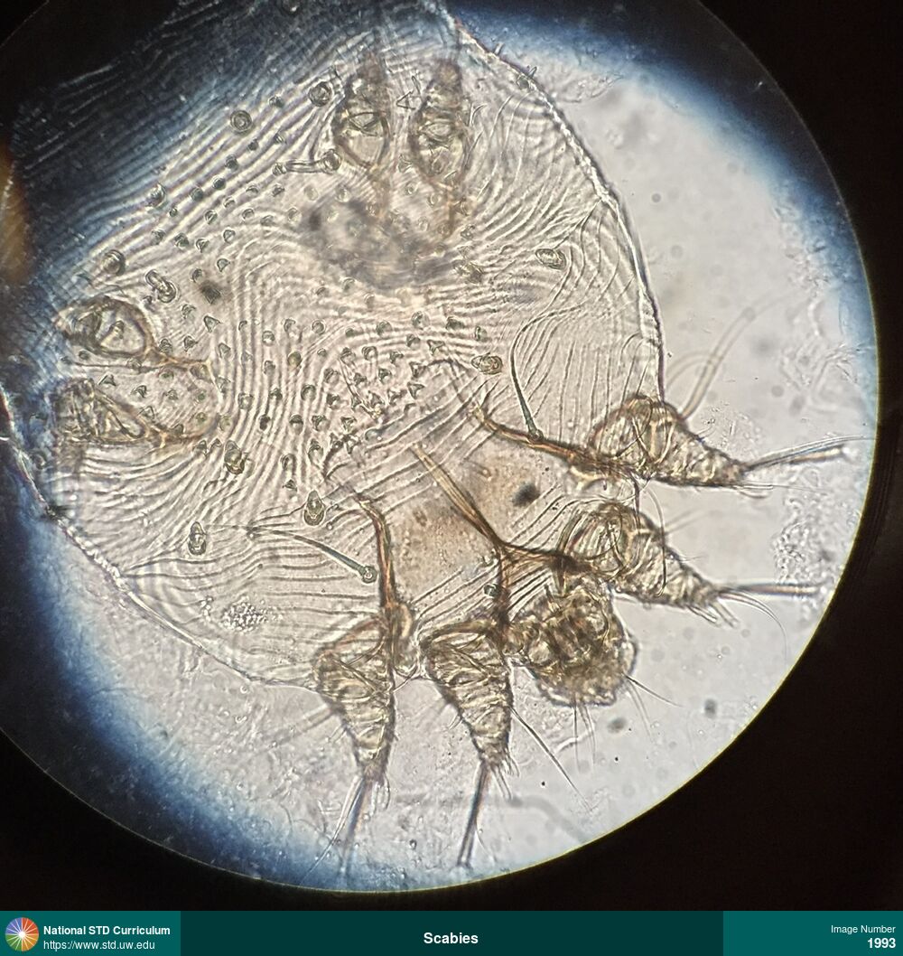
Scabies
Pruritic erythematous papules on right wrist caused by scabies.
Photo: Papule / Papules, Arm (Right), Hand (Right), Itch
Courtesy of Negusse Ocbamichael, PA
Papule / Papules Arm (Right), Hand (Right)
718
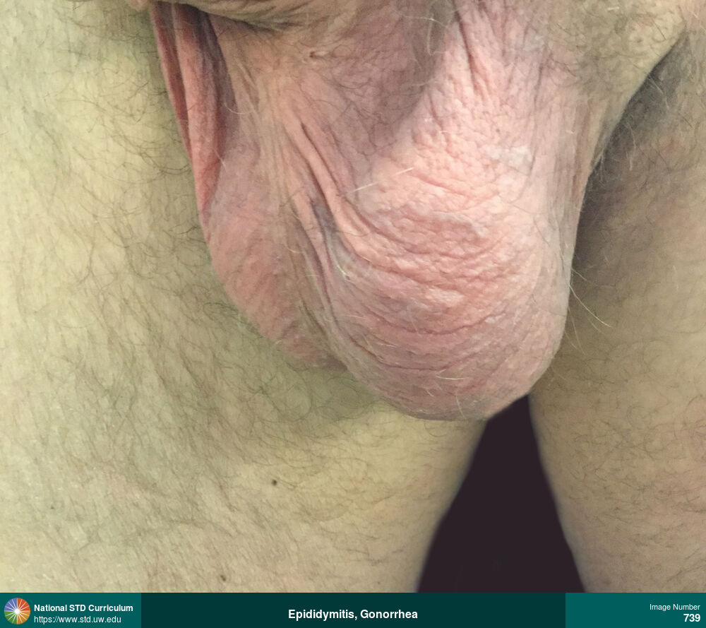
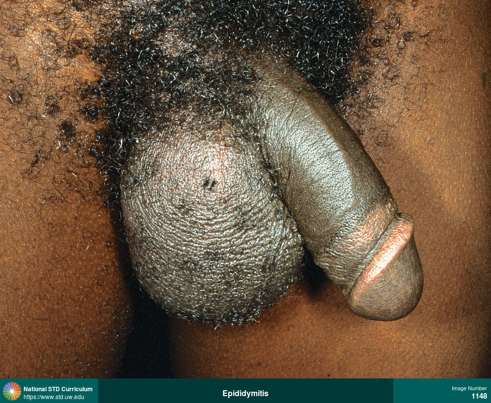
Epididymitis, Gonorrhea
Left-sided testicular and epididymal pain and swelling caused by epididymitis. Urine nucleic acid amplification testing was positive for Neisseria gonorrhoeae.
Photo: Edema / Swelling, Epididymis, Light skin tone, Scrotum, Painful
Courtesy of Negusse Ocbamichael, PA
Edema / Swelling Epididymis, Light skin tone, Scrotum
739

Epididymitis, Gonorrhea
Left-sided testicular and epididymal pain and swelling caused by epididymitis. Urine nucleic acid amplification testing was positive for Neisseria gonorrhoeae.
Photo: Edema / Swelling, Epididymis, Light skin tone, Scrotum, Painful
Courtesy of Negusse Ocbamichael, PA
Edema / Swelling Epididymis, Light skin tone, Scrotum
745

Warts (Anogenital)
Papular genital warts clustered on the left side and septum (midline) of the scrotum.
Photo: Papule / Papules, Verrucous, Dark skin tone, Penis, Scrotum
Courtesy of Negusse Ocbamichael, PA
Papule / Papules, Verrucous Dark skin tone, Penis, Scrotum
760
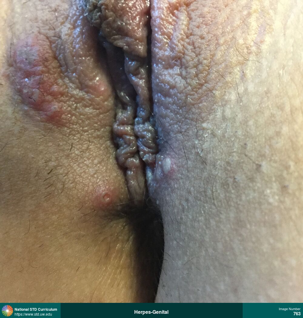
Herpes-Genital
Multiple painful vesicles and ulcers with an erythematous and edematous base on the right labia caused by herpes simplex virus (HSV) infection.
Photo: Edema / Swelling, Erythema, Ulcer / Ulcers, Vesicle / Vesicles, Dark skin tone, Vulva, Painful
Courtesy of Negusse Ocbamichael, PA
Edema / Swelling, Erythema, Ulcer / Ulcers, Vesicle / Vesicles Dark skin tone, Vulva
763
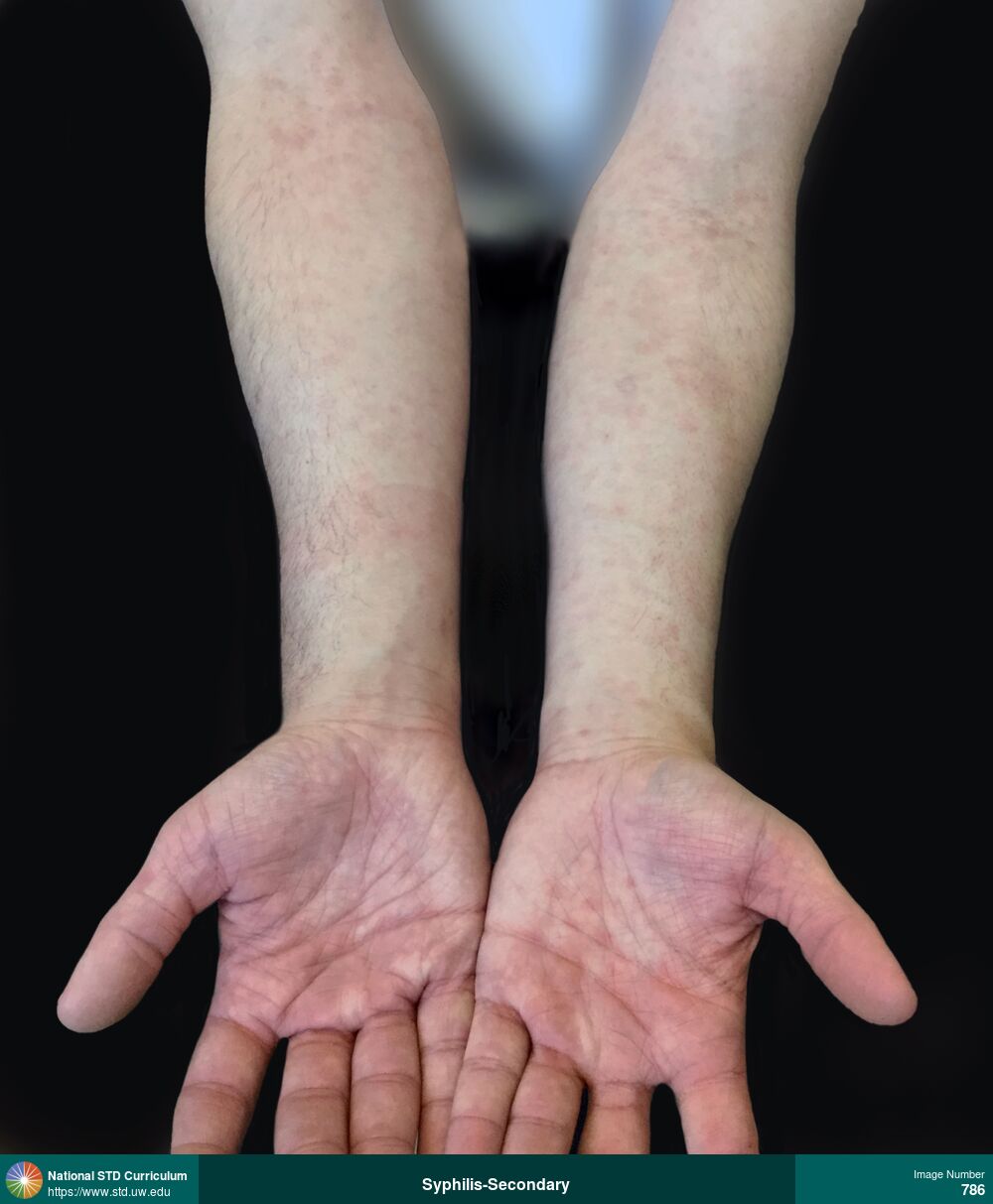
Syphilis-Secondary
Diffuse, erythematous macular rash as shown on on arms and palms in a man with secondary syphilis.
Photo: Erythema, Macule / Macules, Rash, Arm (Left), Arm (Right), Hand (Left), Hand (Right), Rash
Courtesy of Negusse Ocbamichael, PA
Erythema, Macule / Macules, Rash Arm (Left), Arm (Right), Hand (Left), Hand (Right)
786
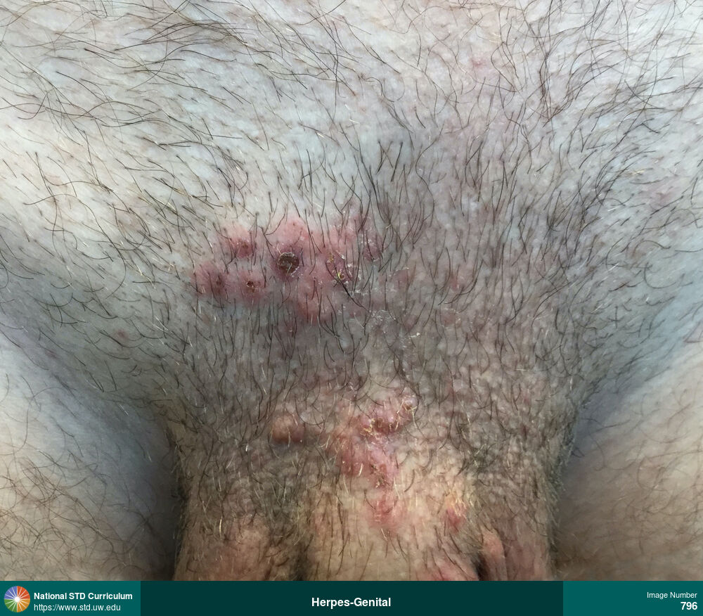
Herpes-Genital
Group of vesicles and shallow ulcers with surrounding erythema located on the base of his penis and suprapubic region in a man with genital herpes simplex virus (HSV) infection. Several of the lesions in the suprapubic regions have formed a scab as part of the healing process.
Photo: Lesion, Scab, Ulcer / Ulcers, Vesicle / Vesicles, Light skin tone, Penis, Suprapubic (Hypogastrium), Painful
Courtesy of Negusse Ocbamichael, PA
Lesion, Scab, Ulcer / Ulcers, Vesicle / Vesicles Light skin tone, Penis, Suprapubic (Hypogastrium)
796

Herpes-Genital
Painful small, tender, ulcers on the shaft and base of the penis in a man with herpes simplex virus type 1 (HSV-1) infection.
Photo: Scab, Ulcer / Ulcers, Vesicle / Vesicles, Light skin tone, Penis, Painful
Courtesy of Negusse Ocbamichael, PA
Scab, Ulcer / Ulcers, Vesicle / Vesicles Light skin tone, Penis
818


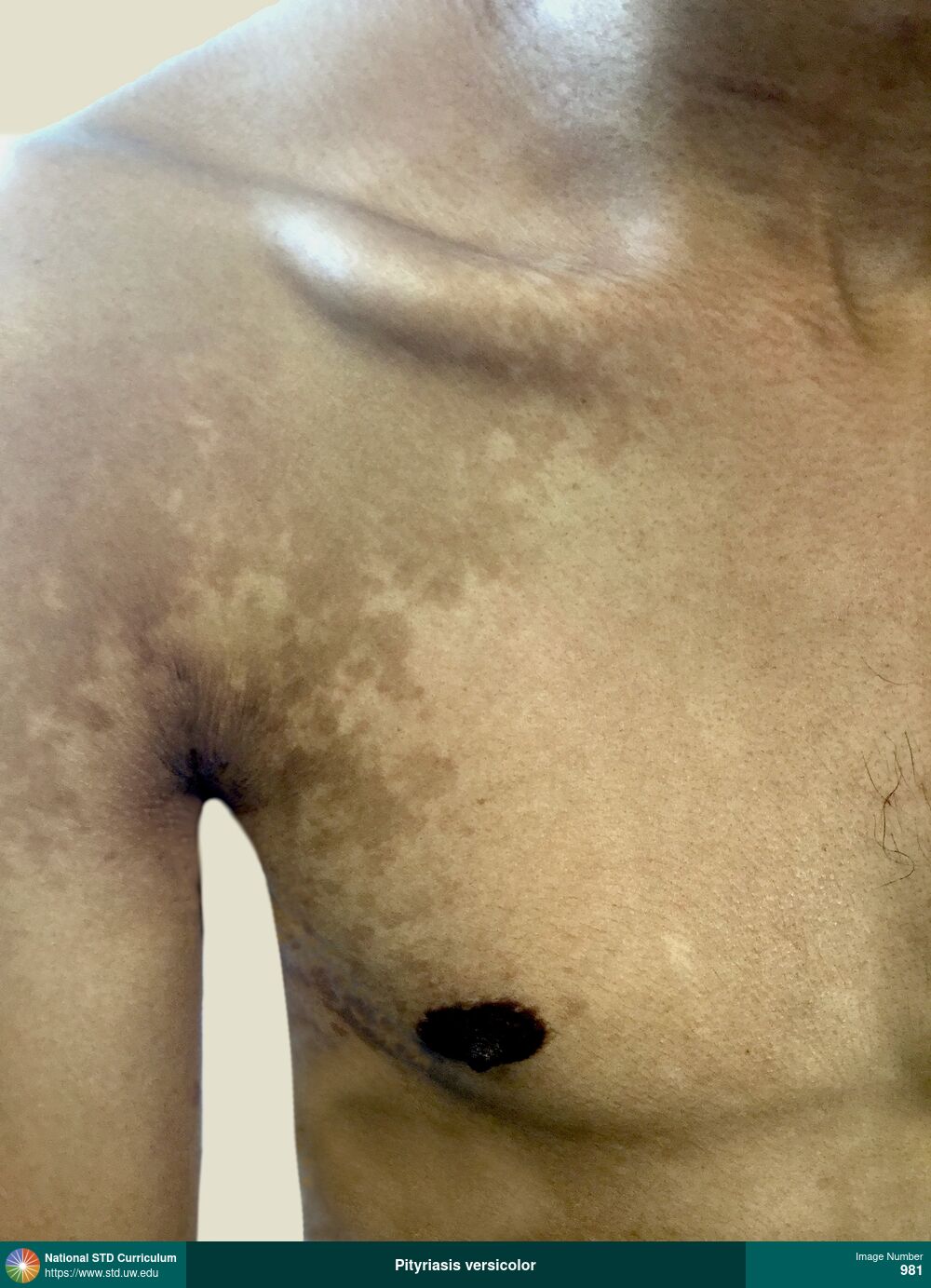
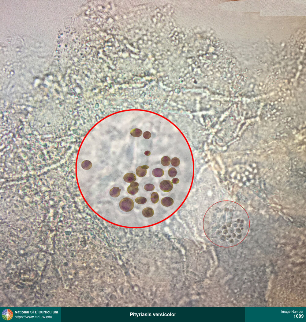
Pityriasis versicolor
Pityriasis versicolor (tinea versicolor) manifesting as hyperpigmented, macular lesions on the chest, shoulders, and back. Note: the involved patches of skin are darker than the surrounding background skin. This condition is caused by overgrowth on the skin with Malassezia yeasts.
Photo: Hyperpigmentation, Macule / Macules, Rash, Chest, Dark skin tone, Shoulder (Right), Itch, Rash
Courtesy of Negusse Ocbamichael, PA
Hyperpigmentation, Macule / Macules, Rash Chest, Dark skin tone, Shoulder (Right)
981

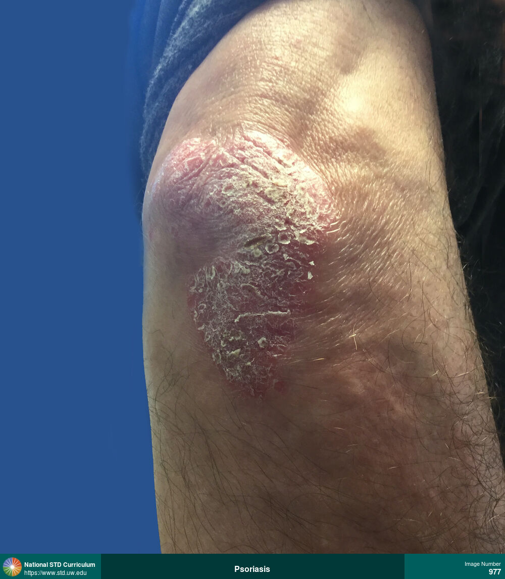
Psoriasis
Psoriasis with erythematous patches on the penis and inguinal region. This type of psoriasis that develops in the genital and inguinal region is referred to as inverse psoriasis, or flexural psoriasis.
Photo: Erythema, Patch/Patches, Rash, Scale, Light skin tone, Penis, Itch
Courtesy of Negusse Ocbamichael, PA
Erythema, Patch/Patches, Rash, Scale Light skin tone, Penis
985
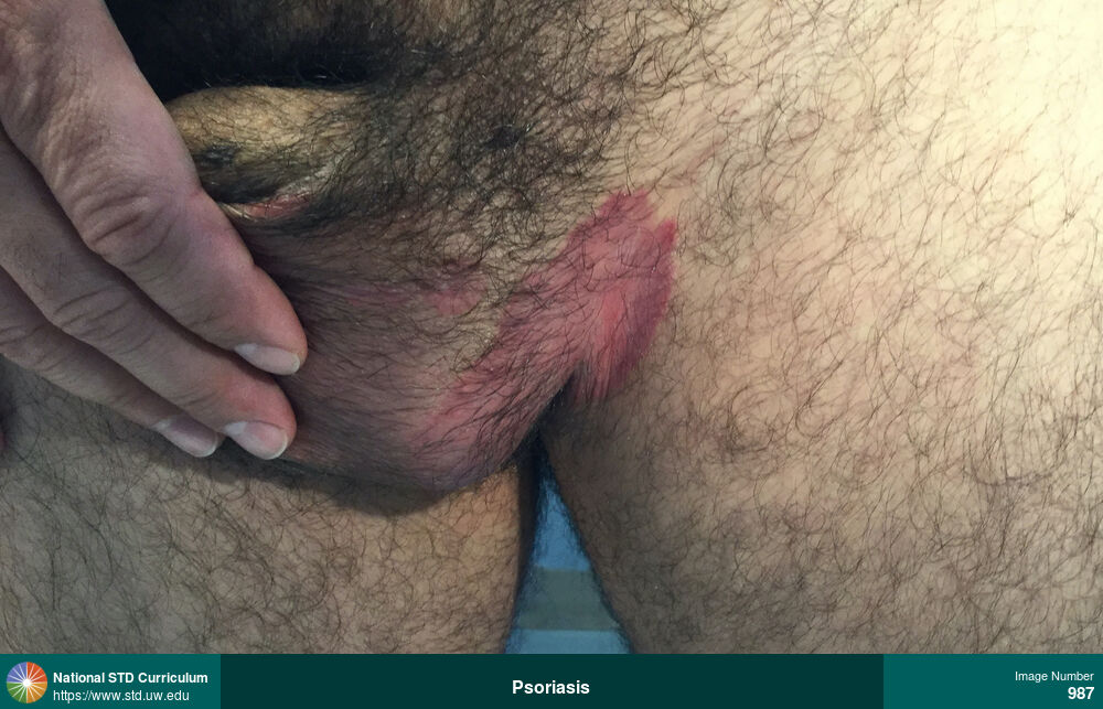
Psoriasis
Psoriasis with erythematous patch in the left inguinal region. This type of psoriasis that develops in the genital and inguinal region is referred to as inverse psoriasis, or flexural psoriasis.
Photo: Erythema, Patch/Patches, Rash, Scale, Groin/Inguinal, Light skin tone, Scrotum, Itch
Courtesy of Negusse Ocbamichael, PA
Erythema, Patch/Patches, Rash, Scale Groin/Inguinal, Light skin tone, Scrotum
987

Psoriasis
Psoriasis with erythematous patch in the bilateral inguinal region. This type of psoriasis that develops in the genital and inguinal region is referred to as inverse psoriasis, or flexural psoriasis.
Photo: Erythema, Patch/Patches, Rash, Scale, Light skin tone, Thigh, Itch
Courtesy of Negusse Ocbamichael, PA
Erythema, Patch/Patches, Rash, Scale Light skin tone, Thigh
989
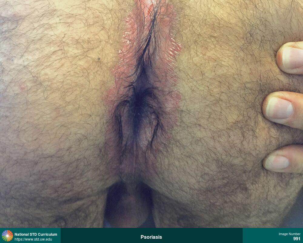
Psoriasis
Psoriasis with erythematous patch in the perianal and medial gluteal region. This type of psoriasis that develops in the genital region is referred to as inverse psoriasis, or flexural psoriasis.
Photo: Erythema, Patch/Patches, Rash, Scale, Anal / Perianal, Buttock, Light skin tone, Itch
Courtesy of Negusse Ocbamichael, PA
Erythema, Patch/Patches, Rash, Scale Anal / Perianal, Buttock, Light skin tone
991
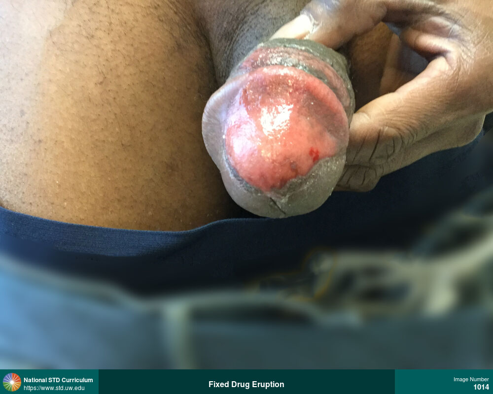
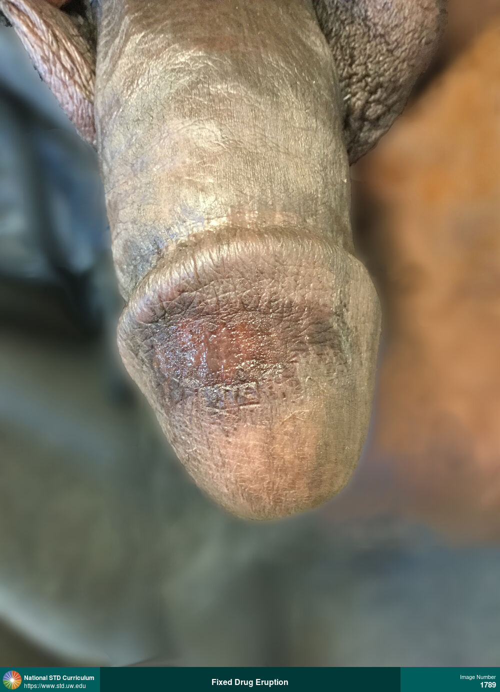
Fixed Drug Eruption
Fixed-drug eruption with a large ulcerated and hypopigmented area on the glans of the penis; the fixed-drug eruption occurred after starting doxycycline for the treatment of urethritis.
Photo: Hypopigmentation, Lesion, Patch/Patches, Ulcer / Ulcers, Dark skin tone, Penis
Courtesy of Negusse Ocbamichael, PA
Hypopigmentation, Lesion, Patch/Patches, Ulcer / Ulcers Dark skin tone, Penis
1014
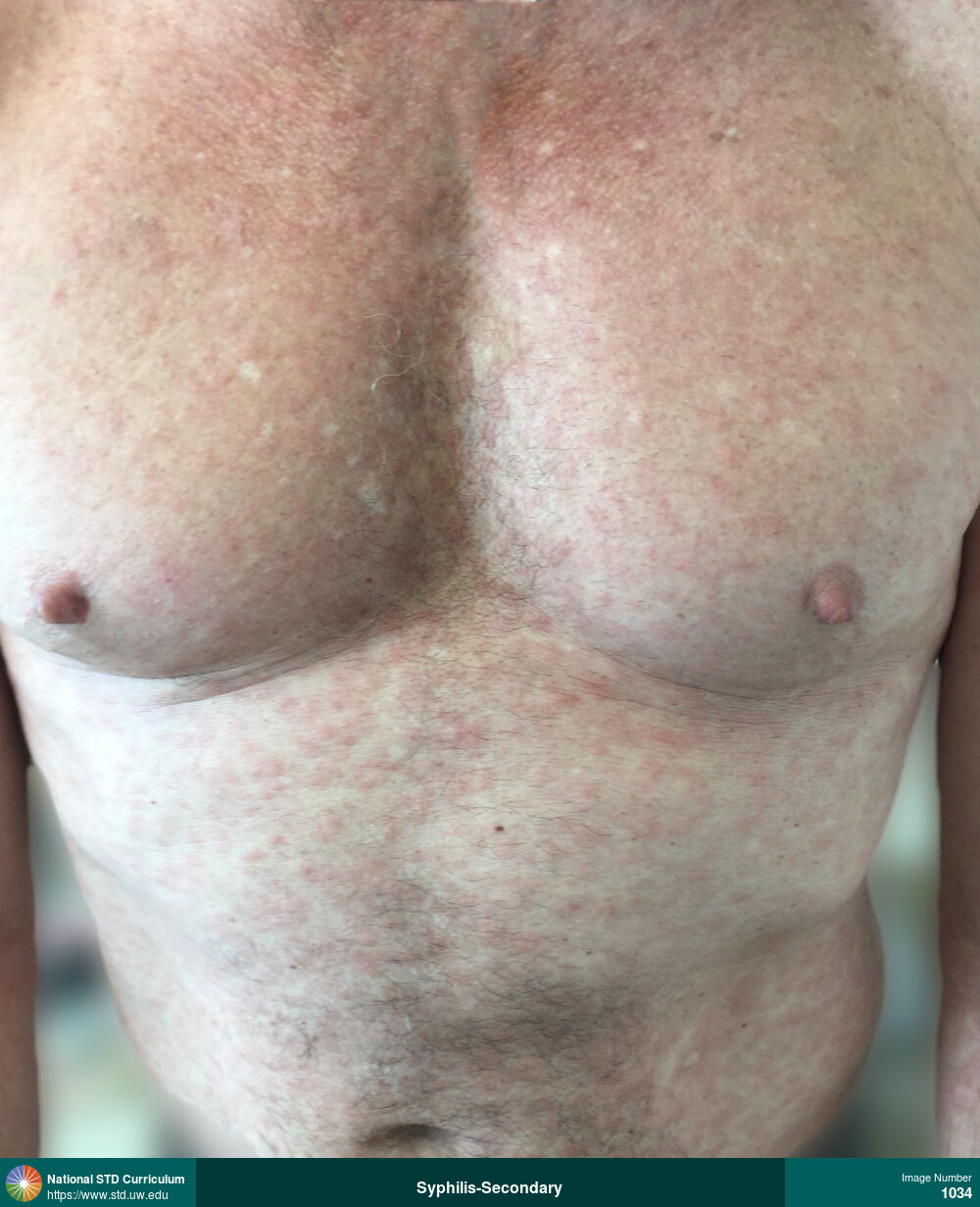
Syphilis-Secondary
Secondary syphilis. Diffuse erythematous Macular rash on body as shown on chest.
Photo: Macule / Macules, Rash, Chest, Light skin tone
Courtesy of Negusse Ocbamichael, PA
Macule / Macules, Rash Chest, Light skin tone
1034
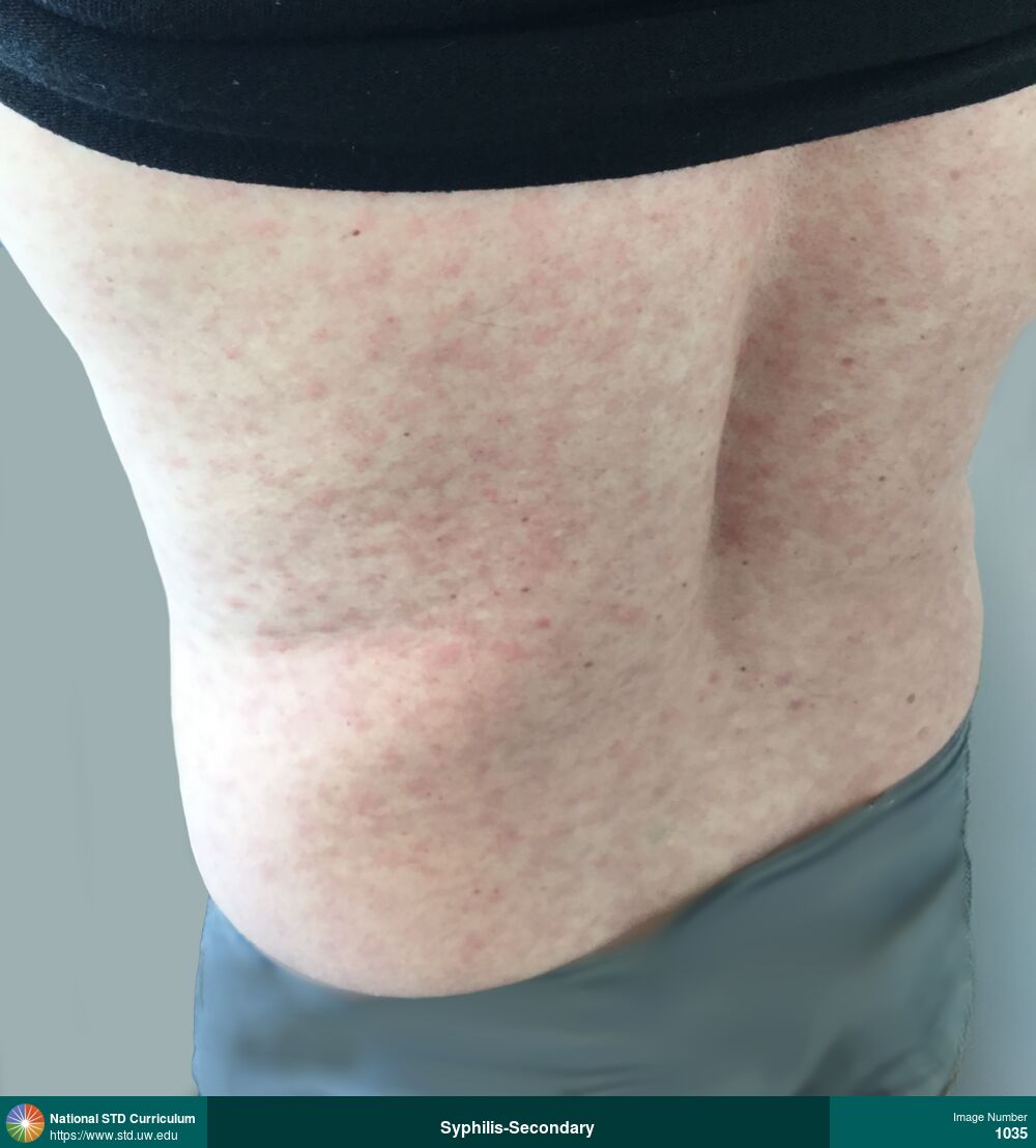
Syphilis-Secondary
Secondary syphilis. Diffuse erythematous Macular rash on body as shown on back.
Photo: Macule / Macules, Rash, Back, Light skin tone
Courtesy of Negusse Ocbamichael, PA
Macule / Macules, Rash Back, Light skin tone
1035
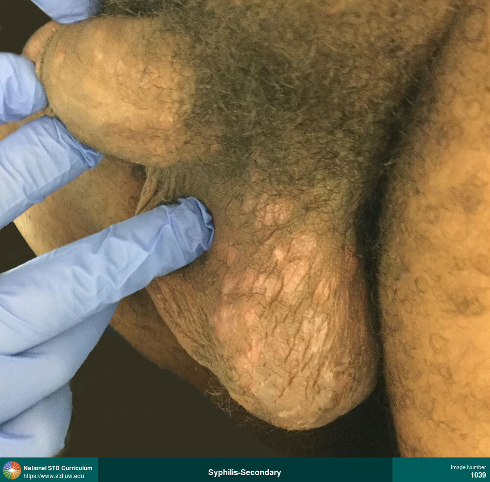
Syphilis-Secondary
Condylomata lata lesions on the scrotum in a man with secondary syphilis.
Photo: Papule / Papules, Plaque, Rash, Dark skin tone, Scrotum
Courtesy of Negusse Ocbamichael, PA
Papule / Papules, Plaque, Rash Dark skin tone, Scrotum
1039
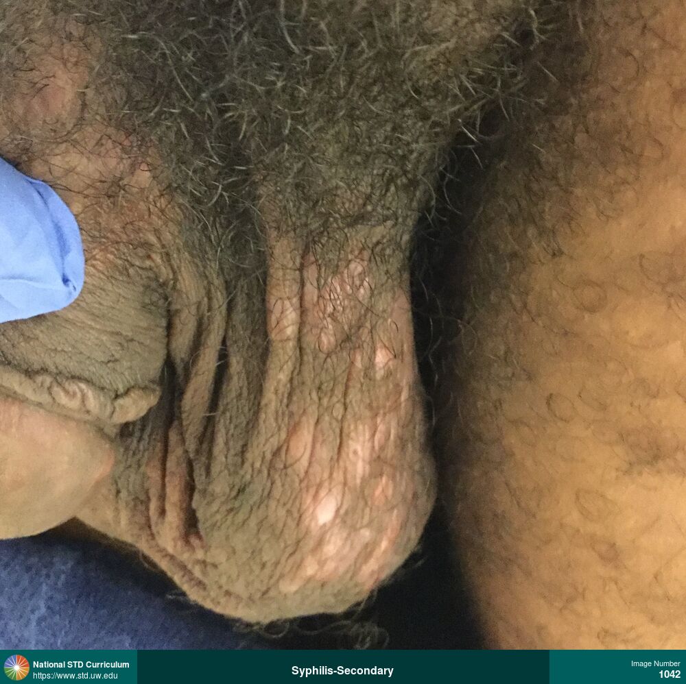
Syphilis-Secondary
Condylomata lata lesions on the scrotum in a man with secondary syphilis.
Photo: Papule / Papules, Plaque, Rash, Dark skin tone, Scrotum
Courtesy of Negusse Ocbamichael, PA
Papule / Papules, Plaque, Rash Dark skin tone, Scrotum
1042

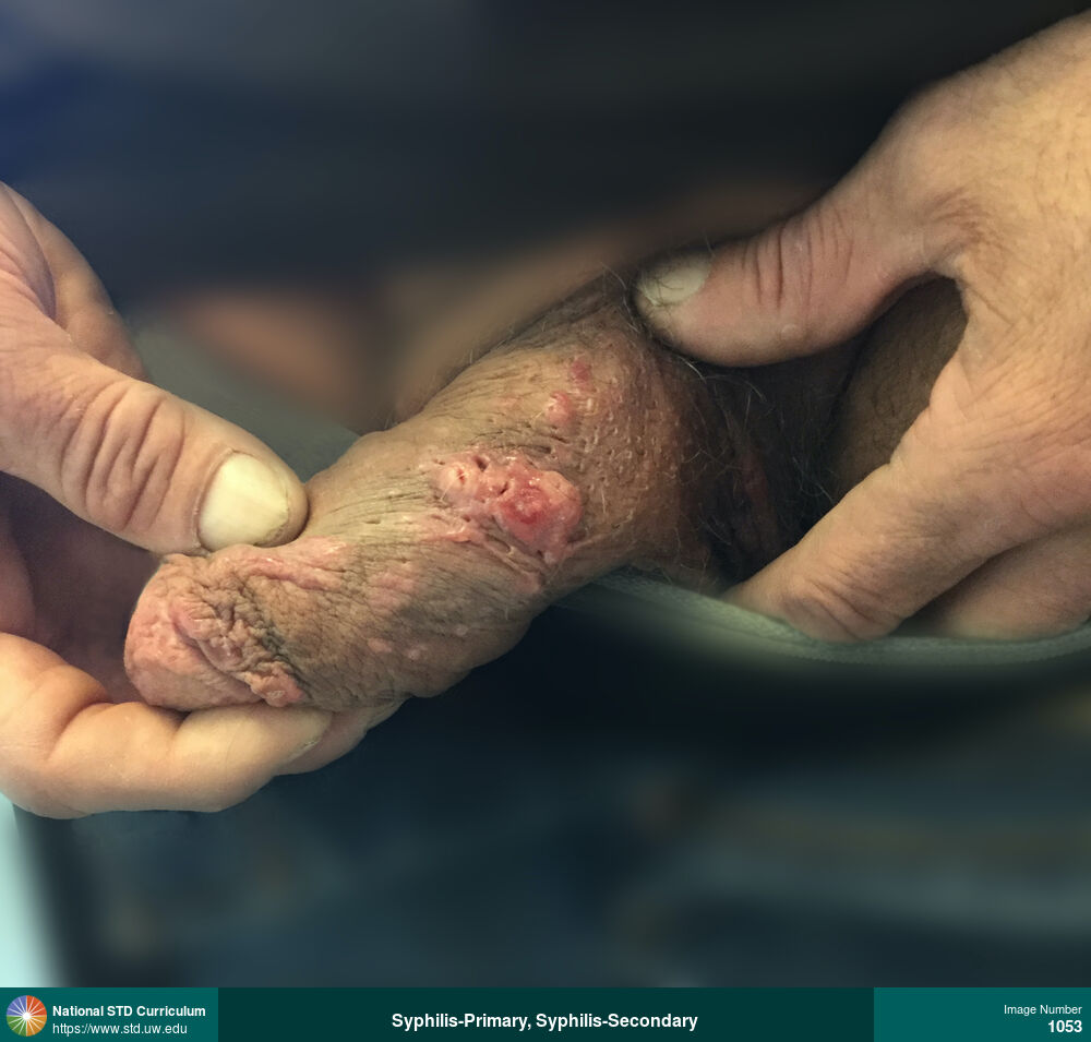
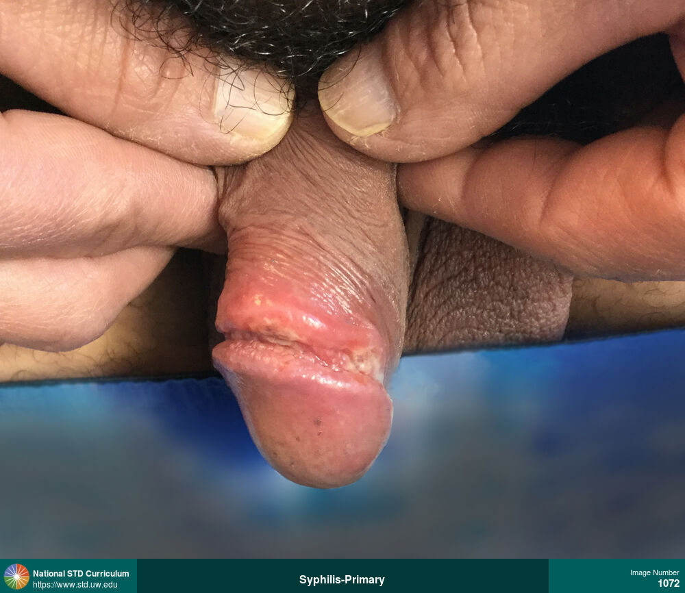
Syphilis-Primary, Syphilis-Secondary
Primary and secondary syphilis. This man had an overlapping penile ulcer (primary syphilis and multiple maculopapular lesions on penis and body (secondary syphilis).
Photo: Lesion, Papule / Papules, Rash, Penis
Courtesy of Negusse Ocbamichael, PA
Lesion, Papule / Papules, Rash Penis
1053
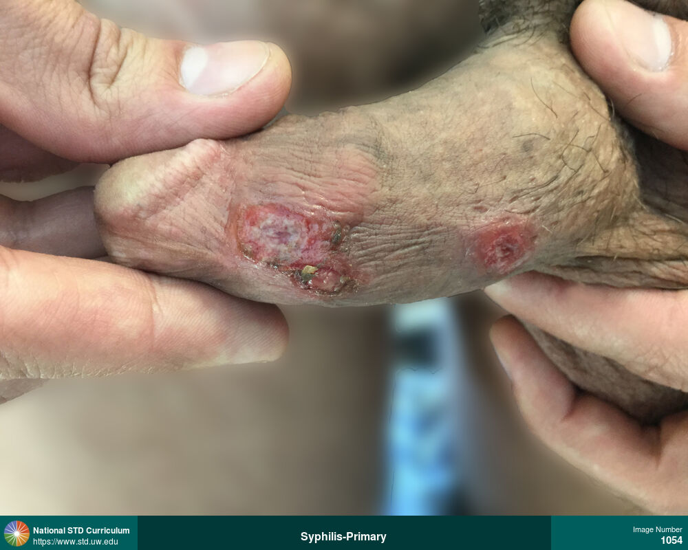
Syphilis-Primary
Photo: Lesion, Ulcer / Ulcers, Penis
Courtesy of Negusse Ocbamichael, PA
Lesion, Ulcer / Ulcers Penis
1054
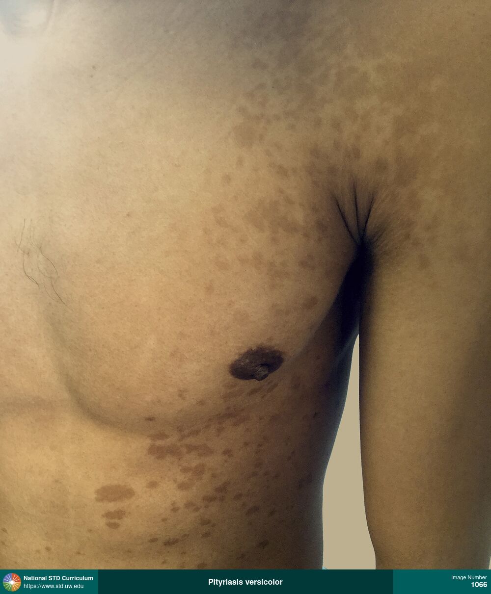
Pityriasis versicolor
Pityriasis versicolor (tinea versicolor) manifesting as hyperpigmented, macular lesions on the chest, shoulders, and back. Note: the involved patches of skin are darker than the surrounding background skin. This condition is caused by overgrowth on the skin with Malassezia yeasts.
Photo: Hyperpigmentation, Macule / Macules, Rash, Chest, Dark skin tone, Shoulder (Left), Itch, Rash
Courtesy of Negusse Ocbamichael, PA
Hyperpigmentation, Macule / Macules, Rash Chest, Dark skin tone, Shoulder (Left)
1066
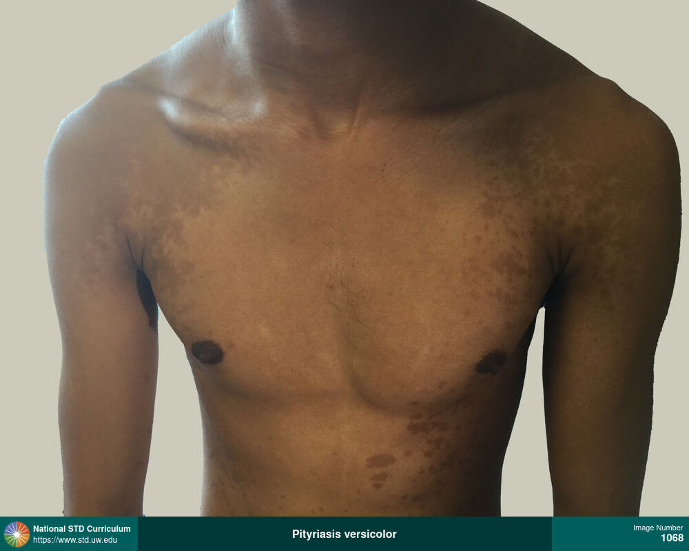
Pityriasis versicolor
Pityriasis versicolor (tinea versicolor) manifesting as hyperpigmented, macular lesions on the chest, shoulders, and back. Note: the involved patches of skin are darker than the surrounding background skin. This condition is caused by overgrowth on the skin with Malassezia yeasts.
Photo: Hyperpigmentation, Macule / Macules, Rash, Chest, Dark skin tone, Itch, Rash
Courtesy of Negusse Ocbamichael, PA
Hyperpigmentation, Macule / Macules, Rash Chest, Dark skin tone
1068
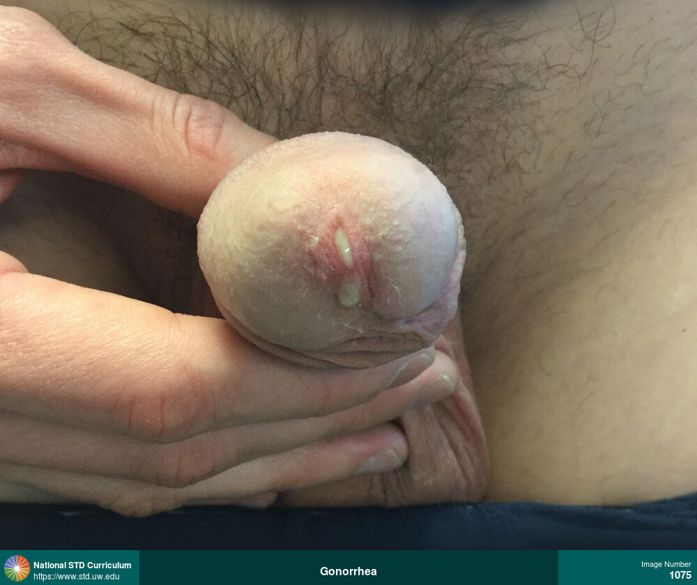
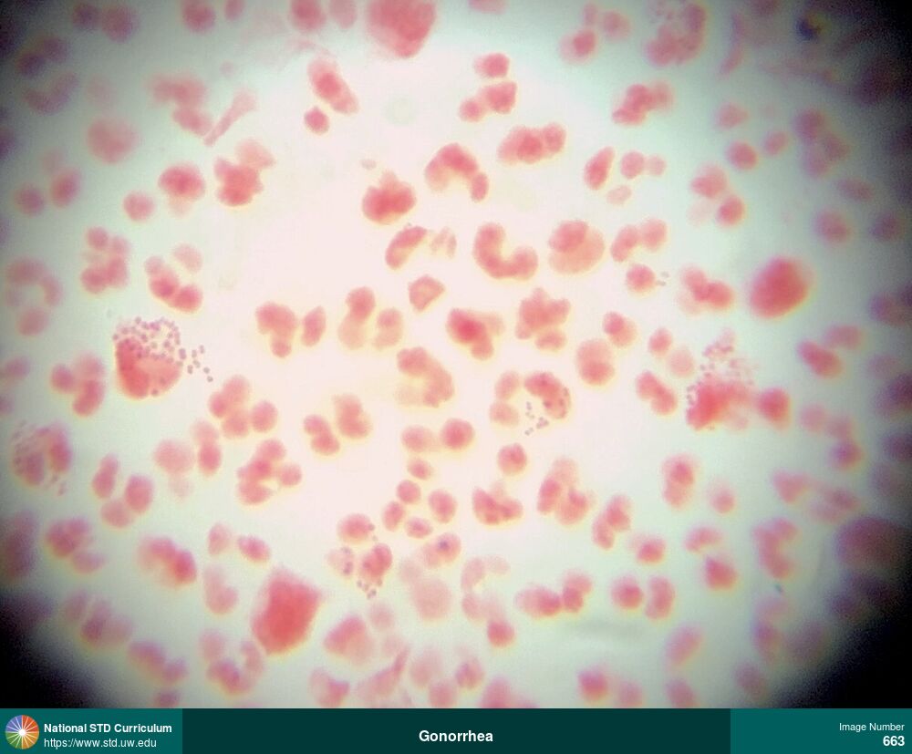
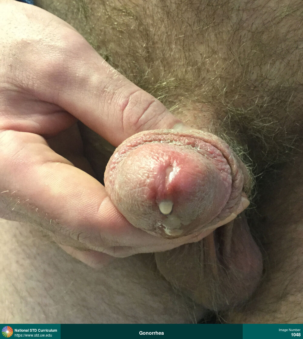
Gonorrhea
Purulent urethral discharge caused by gonorrhea.
Photo: Discharge / Urethra, Light skin tone, Penis
Courtesy of Negusse Ocbamichael, PA
Discharge / Urethra Light skin tone, Penis
1075
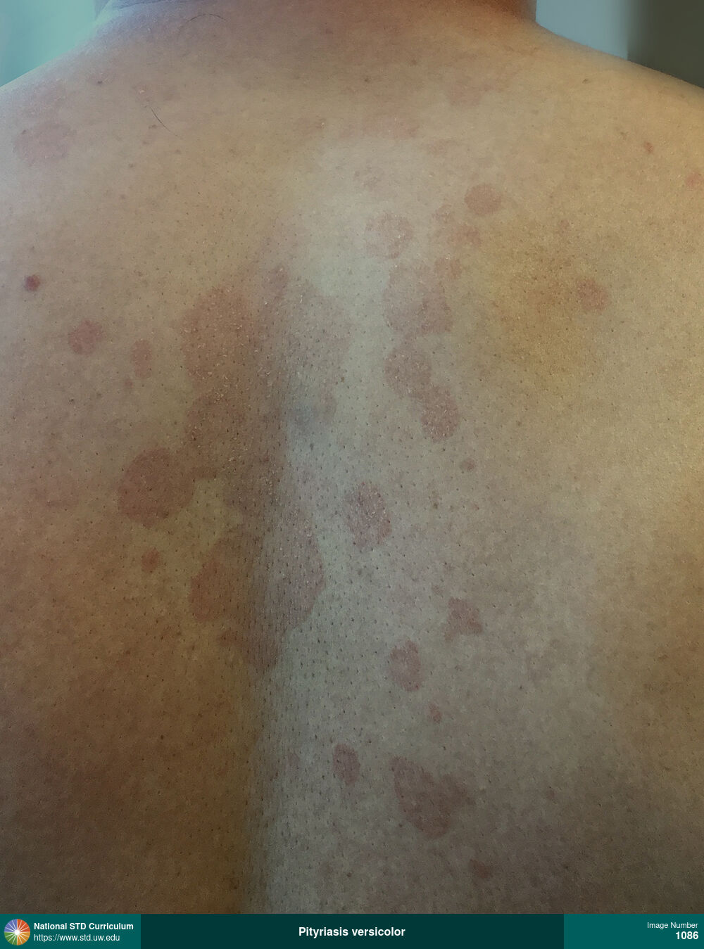
Pityriasis versicolor
Patches of pityriasis versicolor (also referred to as tinea versicolor) on the back. Note: the involved patches of skin are red-brown in color, scaling, and darker than the surrounding unaffected skin.
Photo: Erythema, Hyperpigmentation, Rash, Scale, Back
Courtesy of Negusse Ocbamichael, PA
Erythema, Hyperpigmentation, Rash, Scale Back
1086
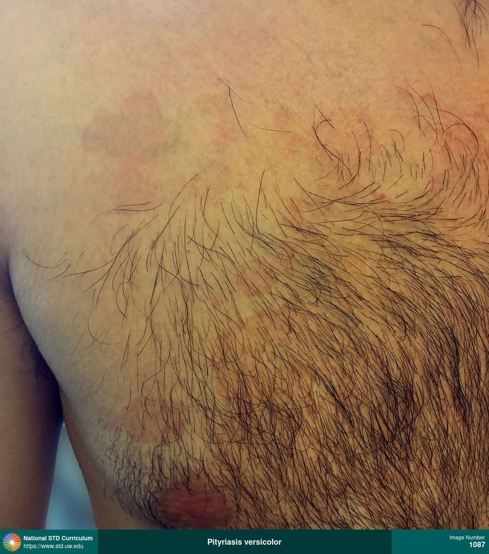
Pityriasis versicolor
Pityriasis versicolor (also known as tinea versicolor) on the right chest region. Note: the involved patches of skin are red-brown in color and more prominent than the surrounding unaffected skin.
Photo: Erythema, Hyperpigmentation, Rash, Chest
Courtesy of Negusse Ocbamichael, PA
Erythema, Hyperpigmentation, Rash Chest
1087
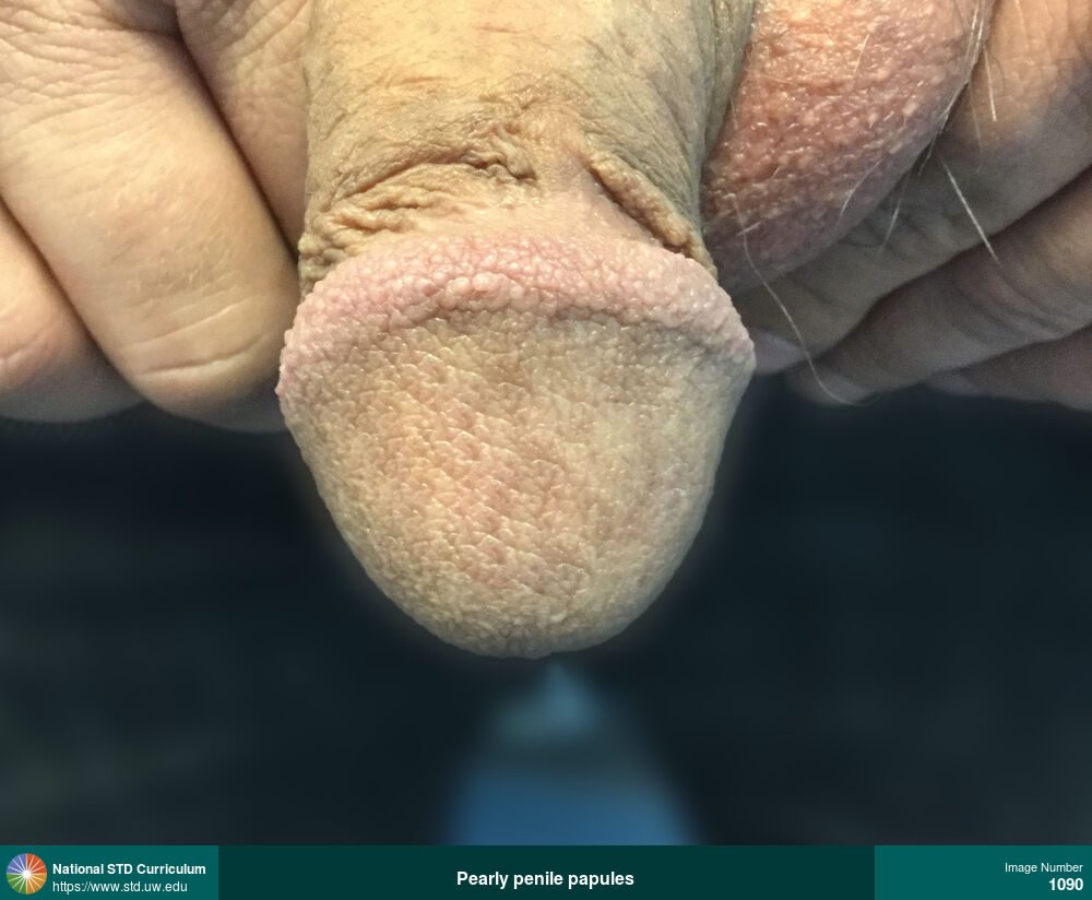
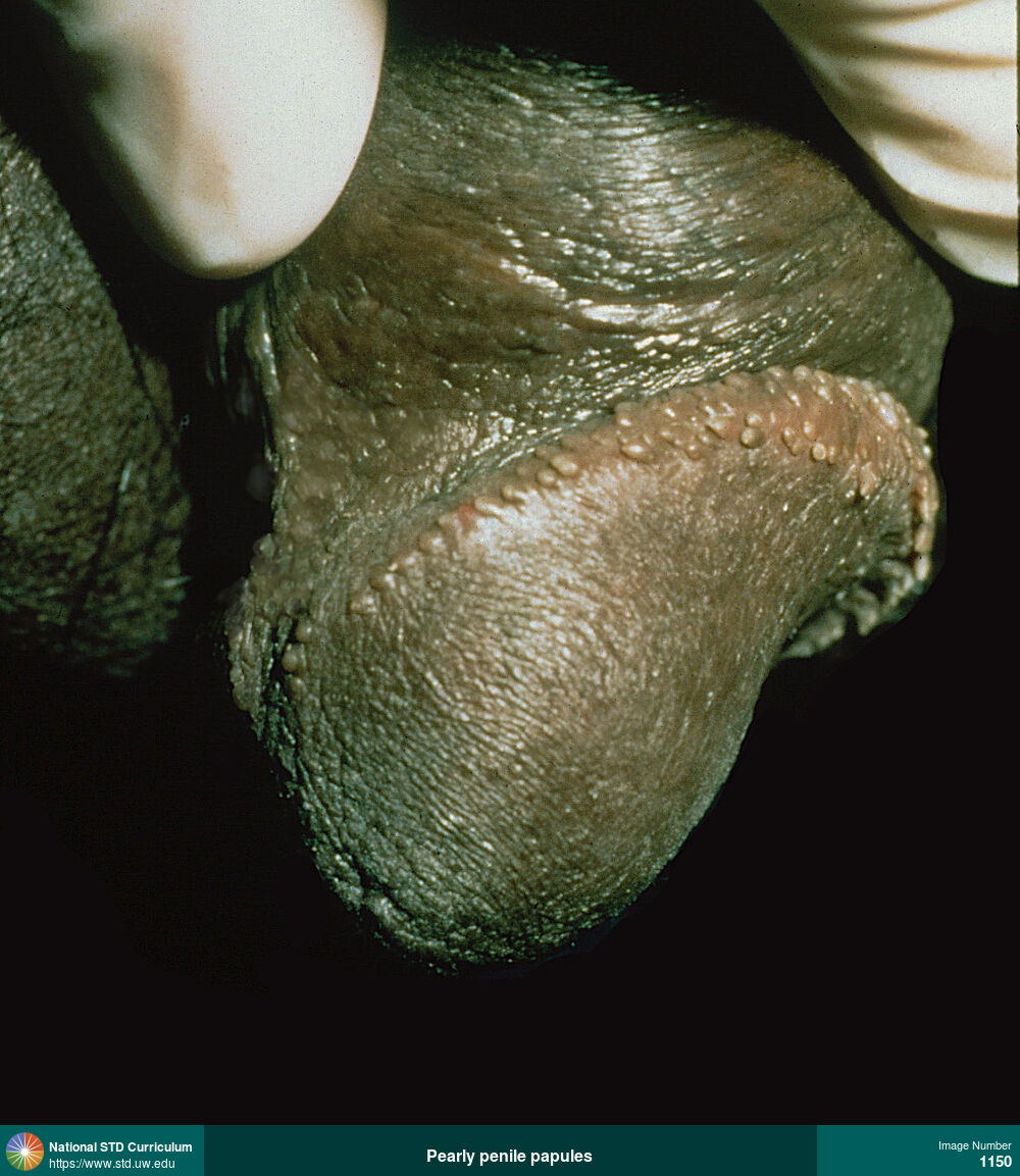
Pearly penile papules
Pearly penile papules are a normal anatomic variant that appear as small (<1 mm), non-tender, flesh-colored bumps in rows around the corona of the penis.
Photo: Papule / Papules, Light skin tone, Penis, Non-Itchy, Non-Painful
Courtesy of Negusse Ocbamichael, PA
Papule / Papules Light skin tone, Penis
1090

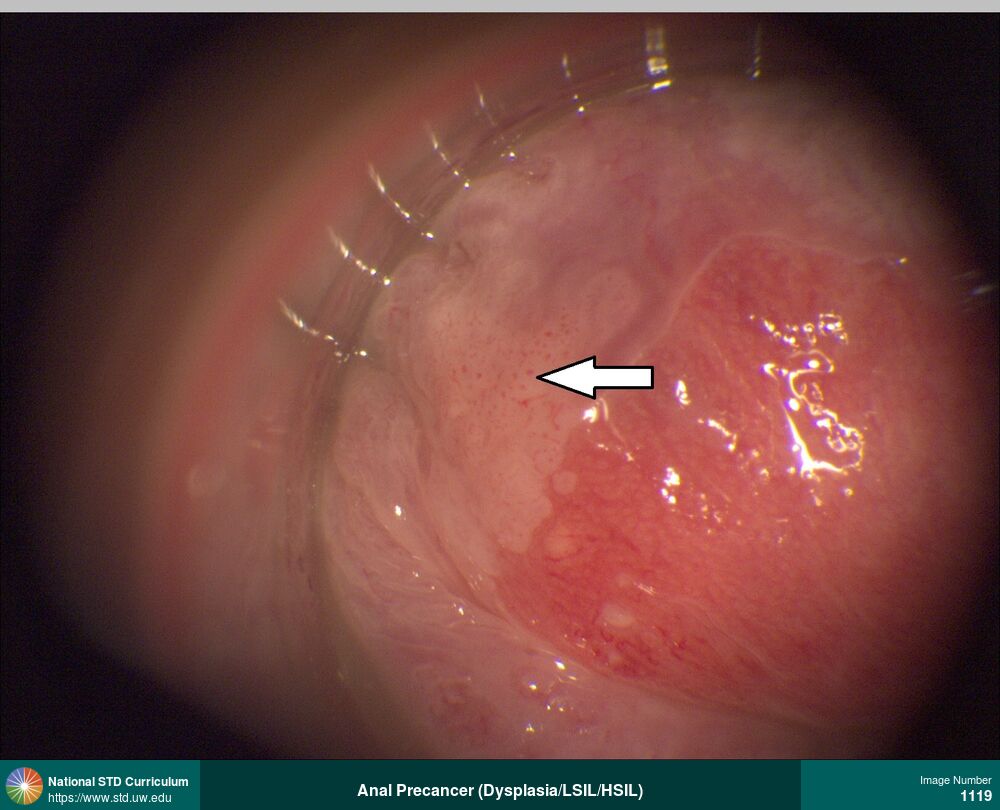
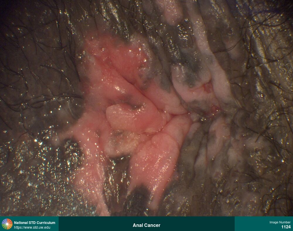
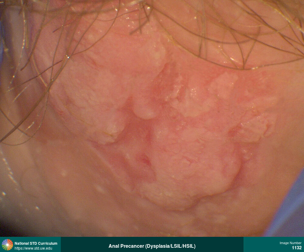
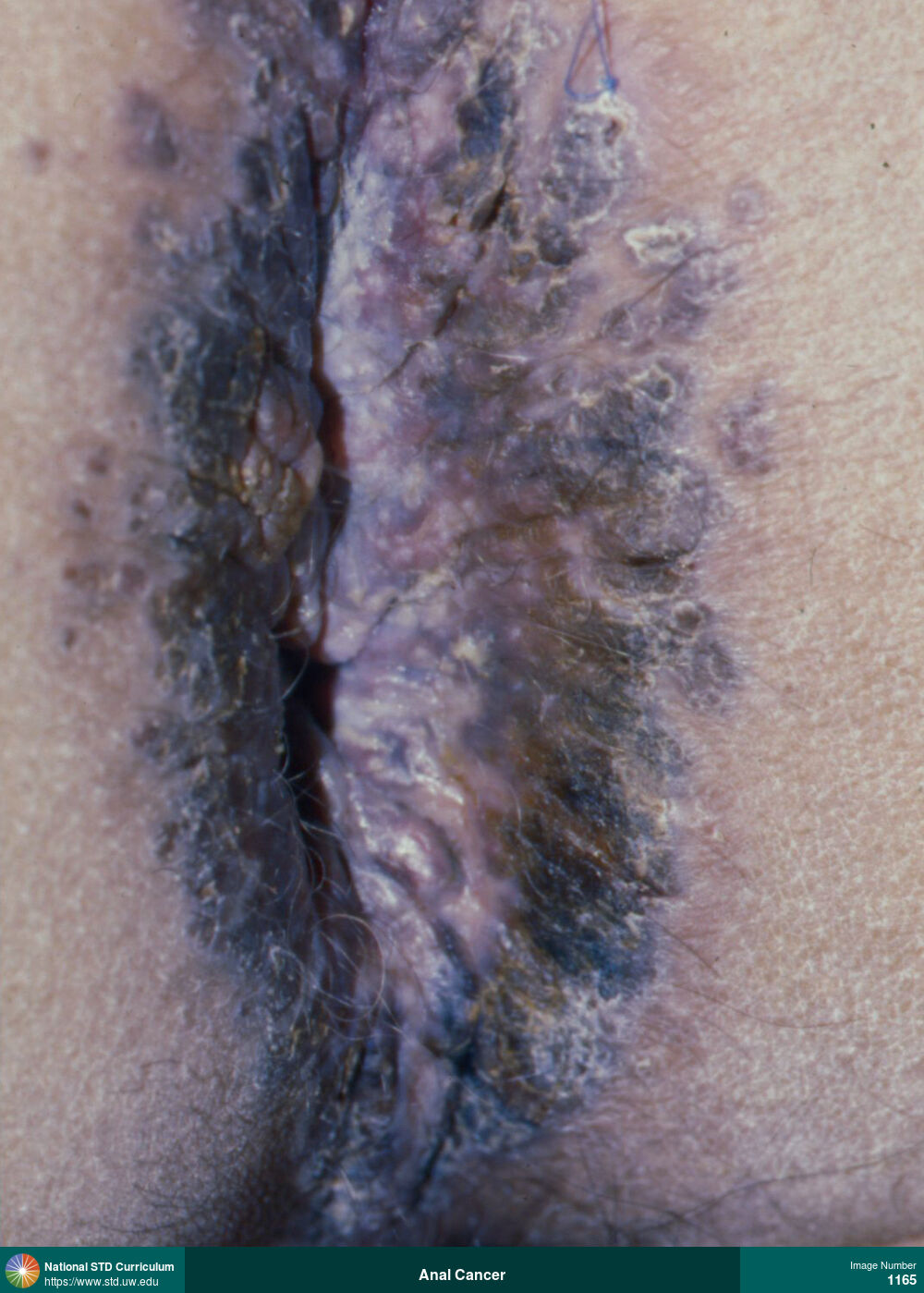
Anal Cancer
Extensive high-grade squamous intraepithelial lesion (HSIL) in perianal region in a man with HIV.
Photo: Lesion, Anal / Intra-anal, Anal / Perianal, Dark skin tone
Courtesy of Jeffrey T. Schouten, MD
Lesion Anal / Intra-anal, Anal / Perianal, Dark skin tone
1124
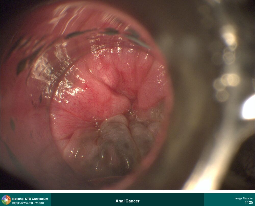
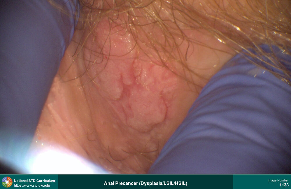
Anal Cancer
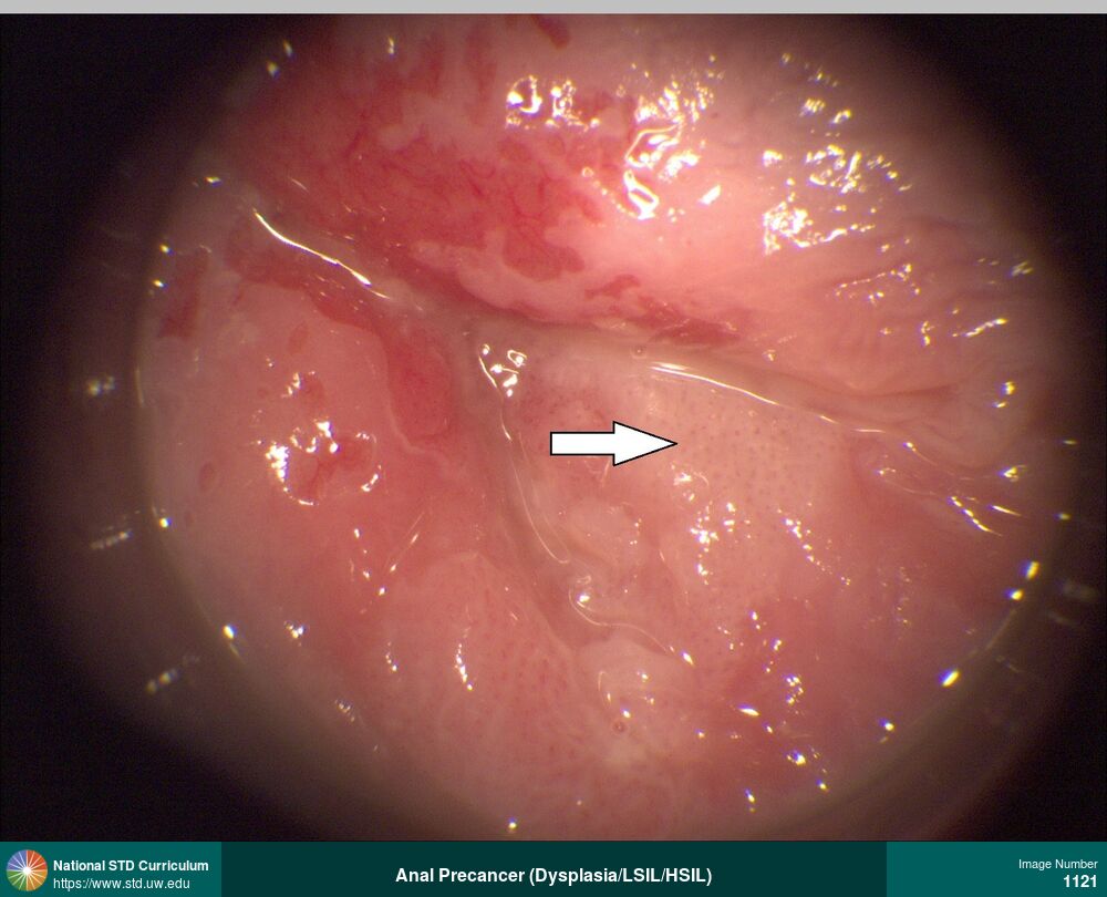
Extensive high-grade squamous intraepithelial lesion (HSIL) in intra-anal region in a man with HIV.
Photo: Anal / Intra-anal, Anal / Perianal, Dark skin tone
Courtesy of Jeffrey T. Schouten, MD
N/A Anal / Intra-anal, Anal / Perianal, Dark skin tone
1125
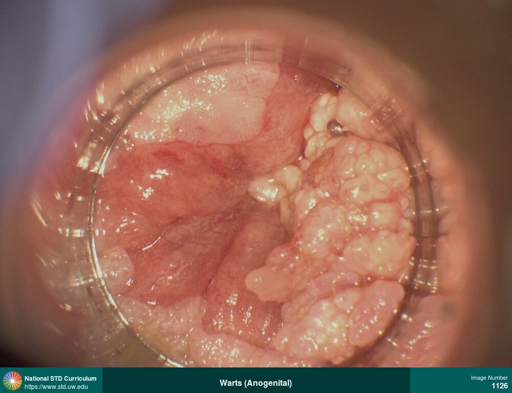
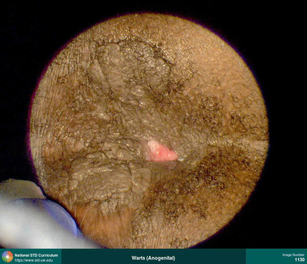
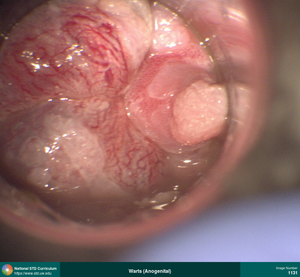
Warts (Anogenital)
Multiple peri-anal condyloma in a man with HIV.
Photo: Papule / Papules, Verrucous, Anal / Perianal, Dark skin tone
Courtesy of Jeffrey T. Schouten, MD
Papule / Papules, Verrucous Anal / Perianal, Dark skin tone
1130
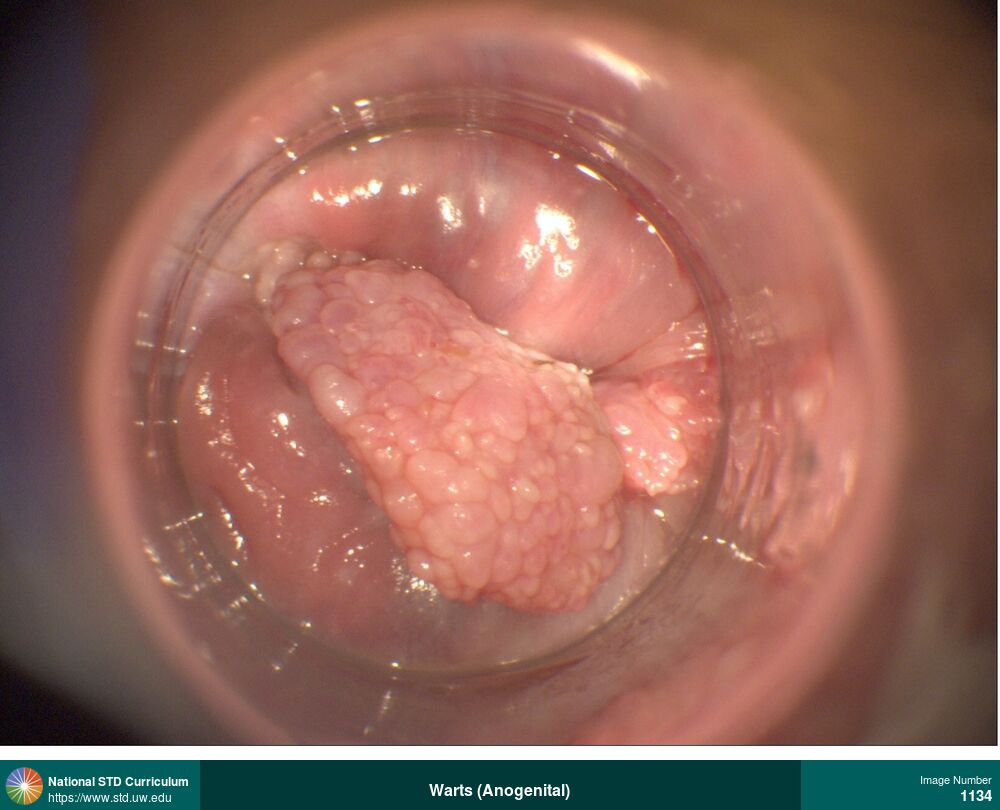
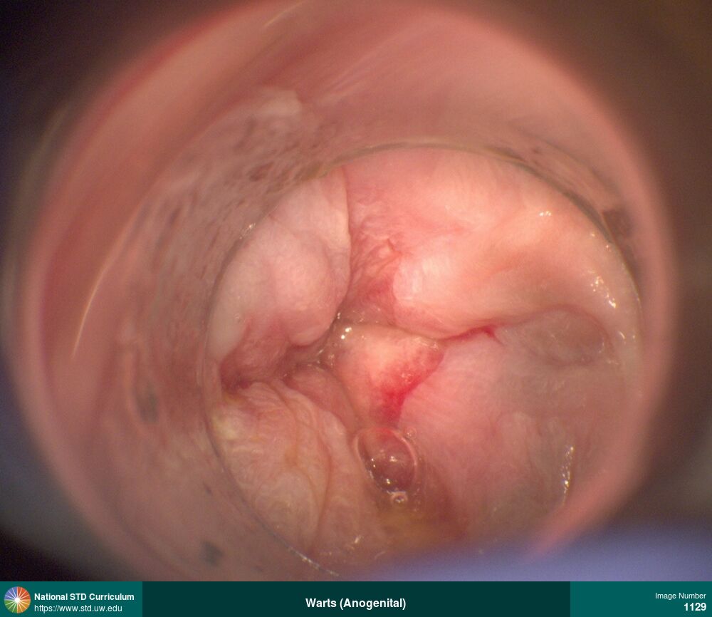
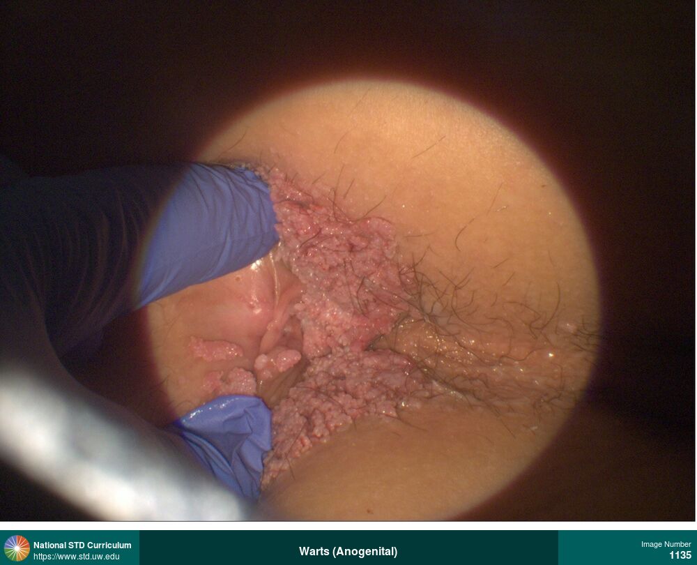
Warts (Anogenital)
Large mass of intra-anal warts in a man with HIV.
Photo: Mass, Verrucous, Anal / Intra-anal
Courtesy of Jeffrey T. Schouten, MD
Mass, Verrucous Anal / Intra-anal
1134
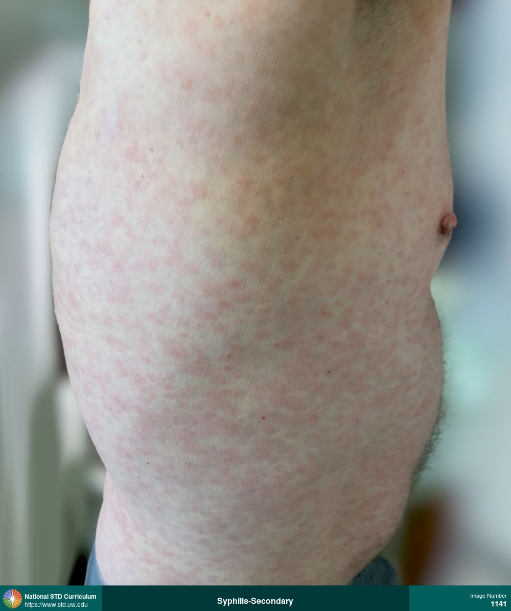
Syphilis-Secondary
Secondary syphilis. Diffuse erythematous Macular rash on body as shown on chest and back (side view).
Photo: Macule / Macules, Rash, Back, Chest, Light skin tone
Courtesy of Negusse Ocbamichael, PA
Macule / Macules, Rash Back, Chest, Light skin tone
1141
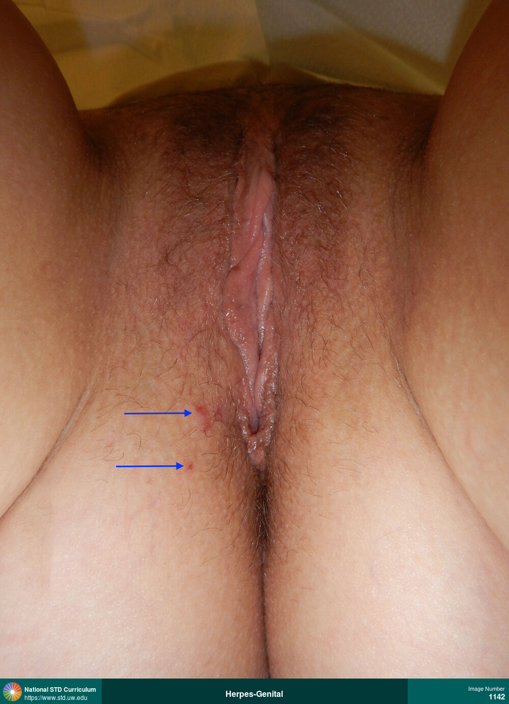
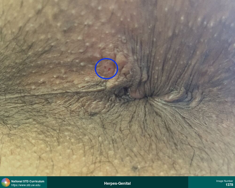
Herpes-Genital
Cluster of superficial ulcerations with slight surrounding erythema present in the lower vaginal/perianal region (blue arrows) caused by recurrent herpes simplex virus (HSV) infection.
Photo: Erythema, Ulcer / Ulcers, Anal / Perianal, Light skin tone, Vulva
Courtesy of Christine M. Johnston, MD, MPH
Erythema, Ulcer / Ulcers Anal / Perianal, Light skin tone, Vulva
1142
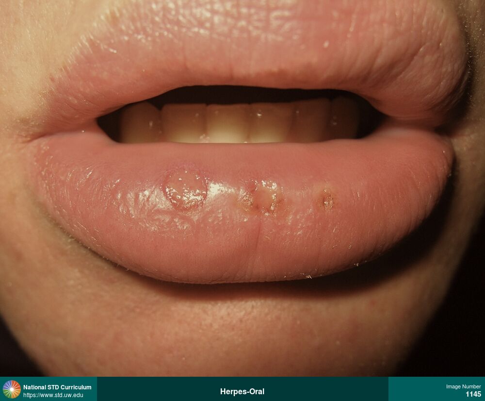
Herpes-Oral
Multiple ulcerated lesions on the lower lip caused by recurrent oral herpes simplex virus (HSV) infection.
Photo: Edema / Swelling, Ulcer / Ulcers, Light skin tone, Lips
Courtesy of Christine M. Johnston, MD, MPH
Edema / Swelling, Ulcer / Ulcers Light skin tone, Lips
1145
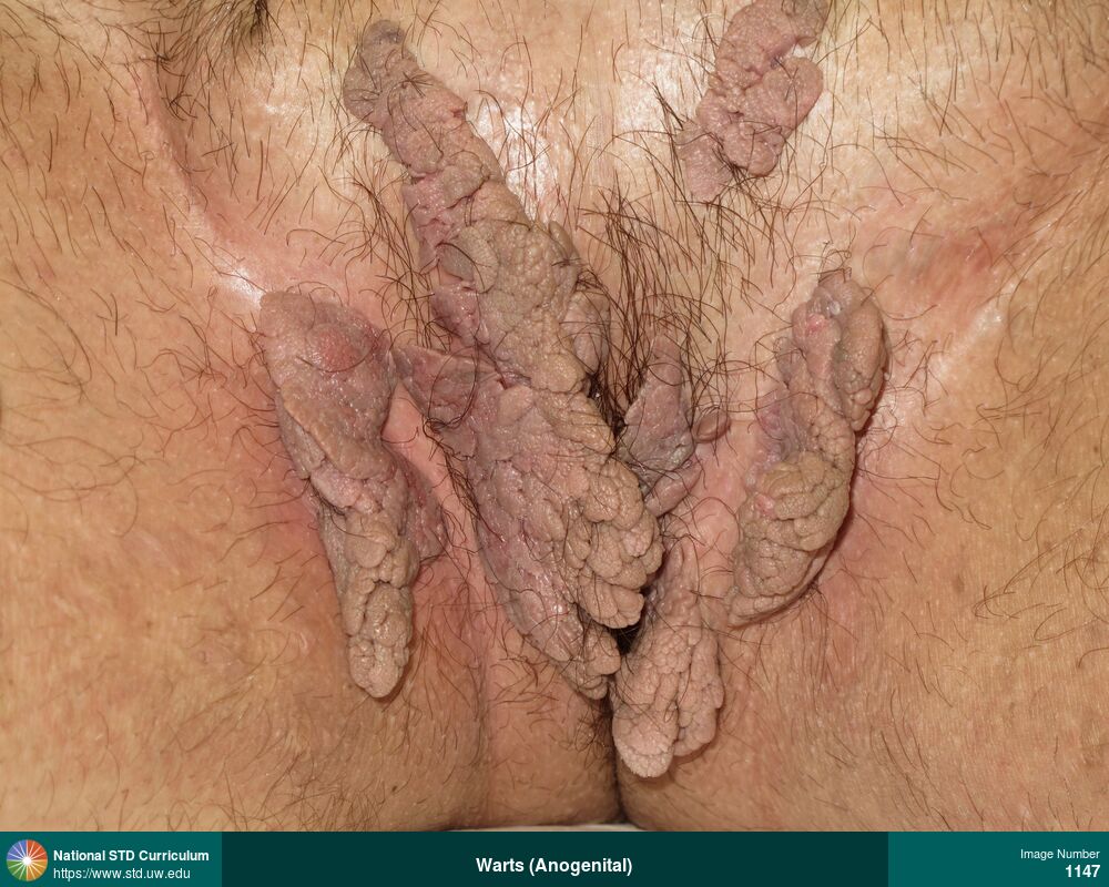
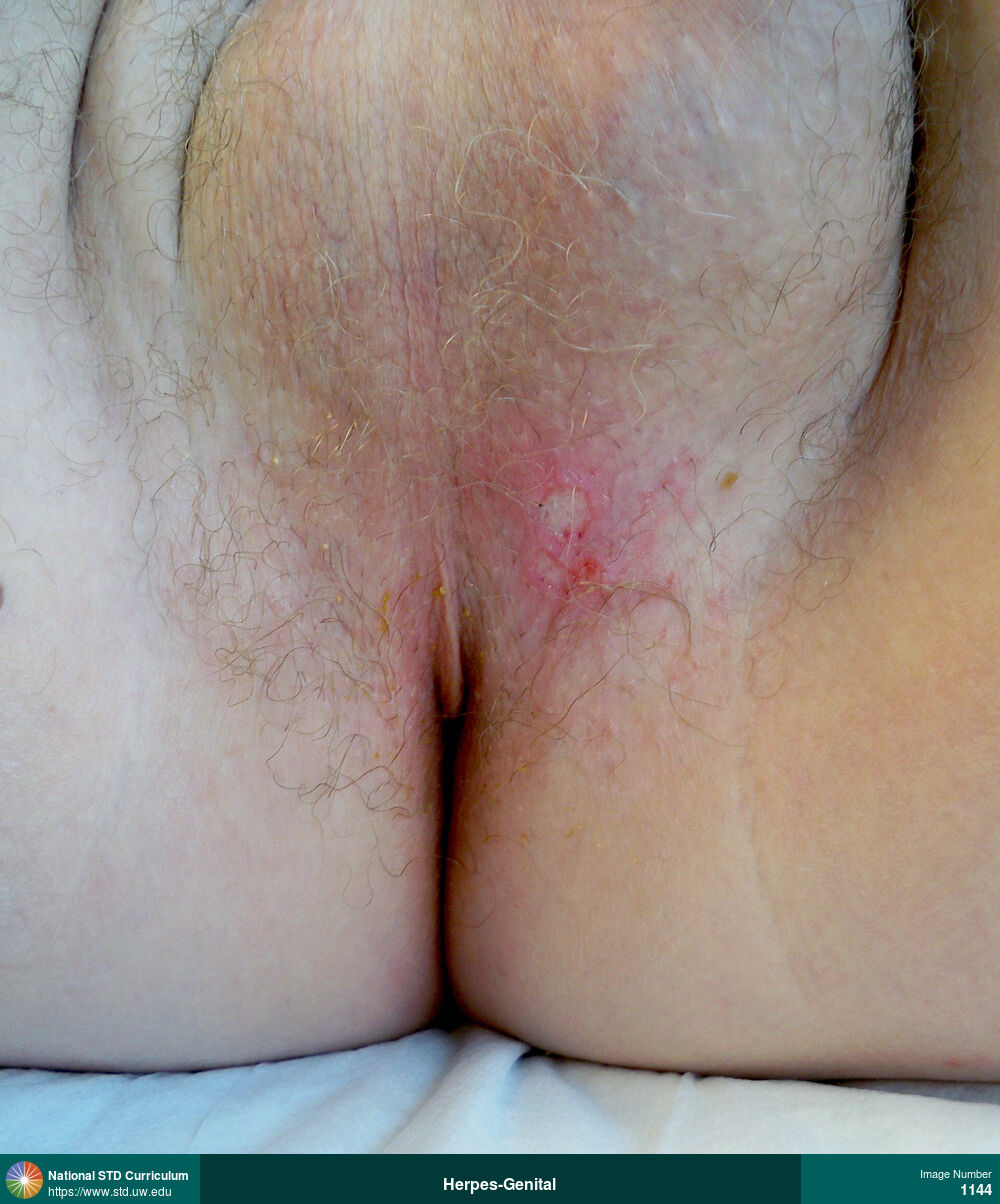
HPV-Related Cancers, Warts (Anogenital)
Extensive perianal warts in a man.
Photo: Verrucous, Anal / Perianal, Groin/Inguinal, Light skin tone, Scrotum, Non-Painful
Courtesy of Seattle & King County Sexual Health Clinic
Verrucous Anal / Perianal, Groin/Inguinal, Light skin tone, Scrotum
1147
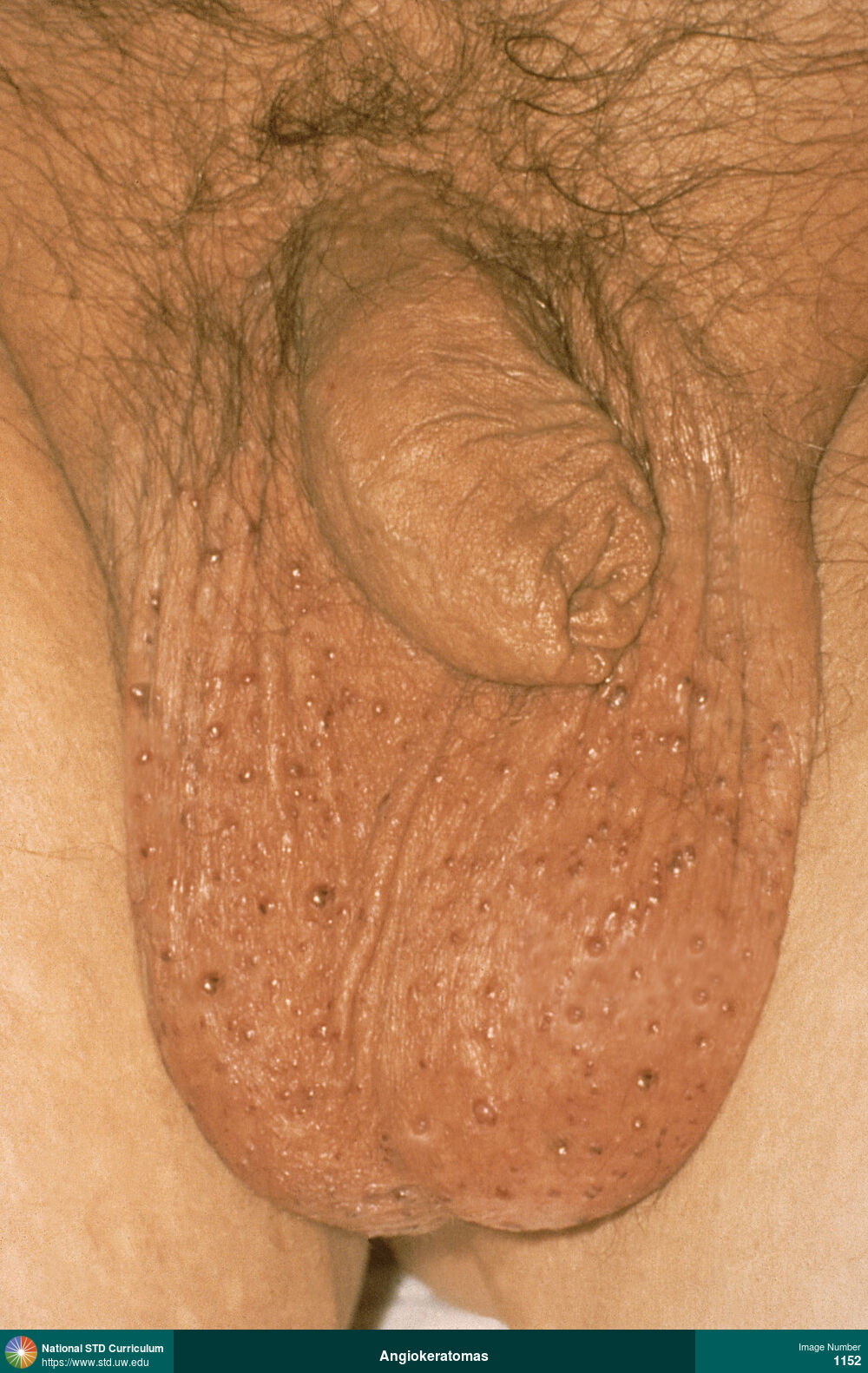
Angiokeratomas
Angiokeratomas (or Fordyce angiokeratomas). This man had chronic multiple small red-dark purple, non-tender papular lesions on the scrotum.
Photo: Papule / Papules, Light skin tone, Scrotum, Non-Painful
Courtesy of Seattle & King County Sexual Health Clinic
Papule / Papules Light skin tone, Scrotum
1152
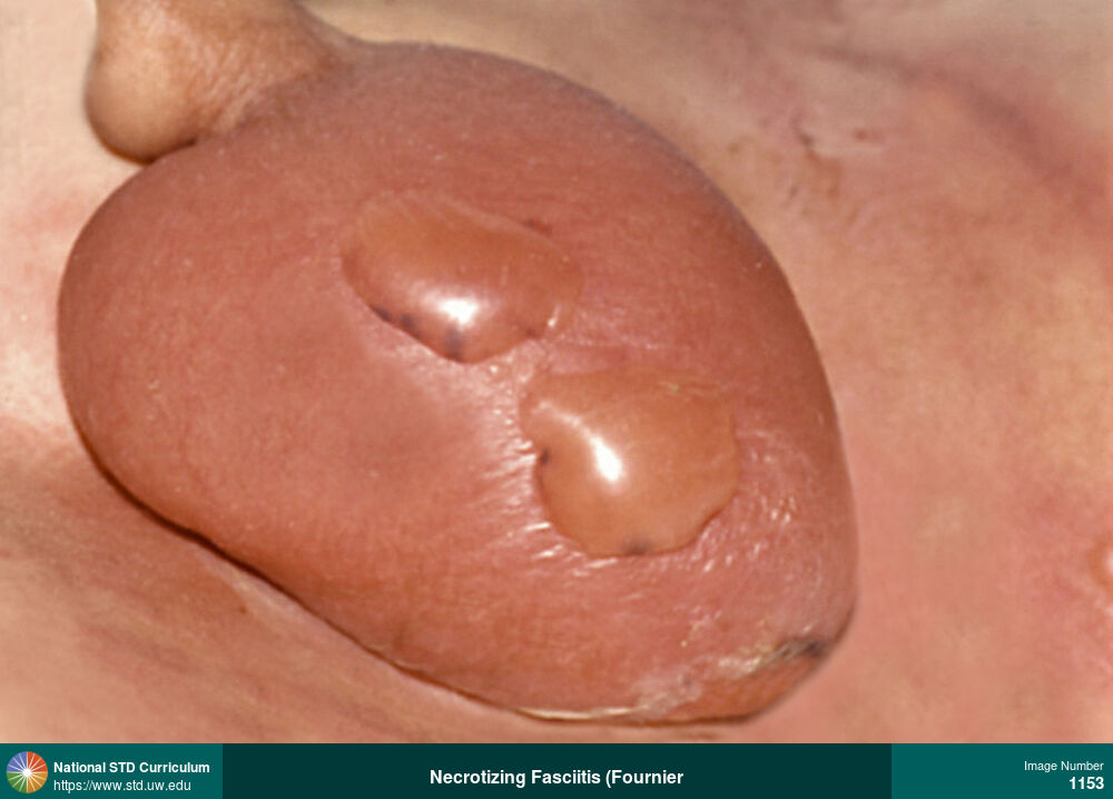
Necrotizing Fasciitis (Fournier's gangrene)
Scrotal cellulitis (with severe edema and bullous lesions) in a man with acute lymphocytic leukemia (ALL) and neutropenia; he was diagnosed with necrotizing fasciitis (Fournier’s gangrene).
Photo: Bulla / Bullae, Edema / Swelling, Erythema, Light skin tone, Scrotum, Painful
Courtesy of David H. Spach, MD
Bulla / Bullae, Edema / Swelling, Erythema Light skin tone, Scrotum
1153
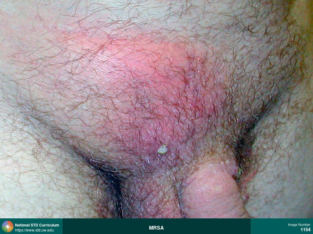
MRSA
Suprapubic abscess and cellulitis caused by methicillin-resistant Staphylococcus aureus (MRSA).
Photo: Abscess, Edema / Swelling, Erythema, Pustule / Pustules, Light skin tone, Suprapubic (Hypogastrium)
Courtesy of David H. Spach, MD
Abscess, Edema / Swelling, Erythema, Pustule / Pustules Light skin tone, Suprapubic (Hypogastrium)
1154
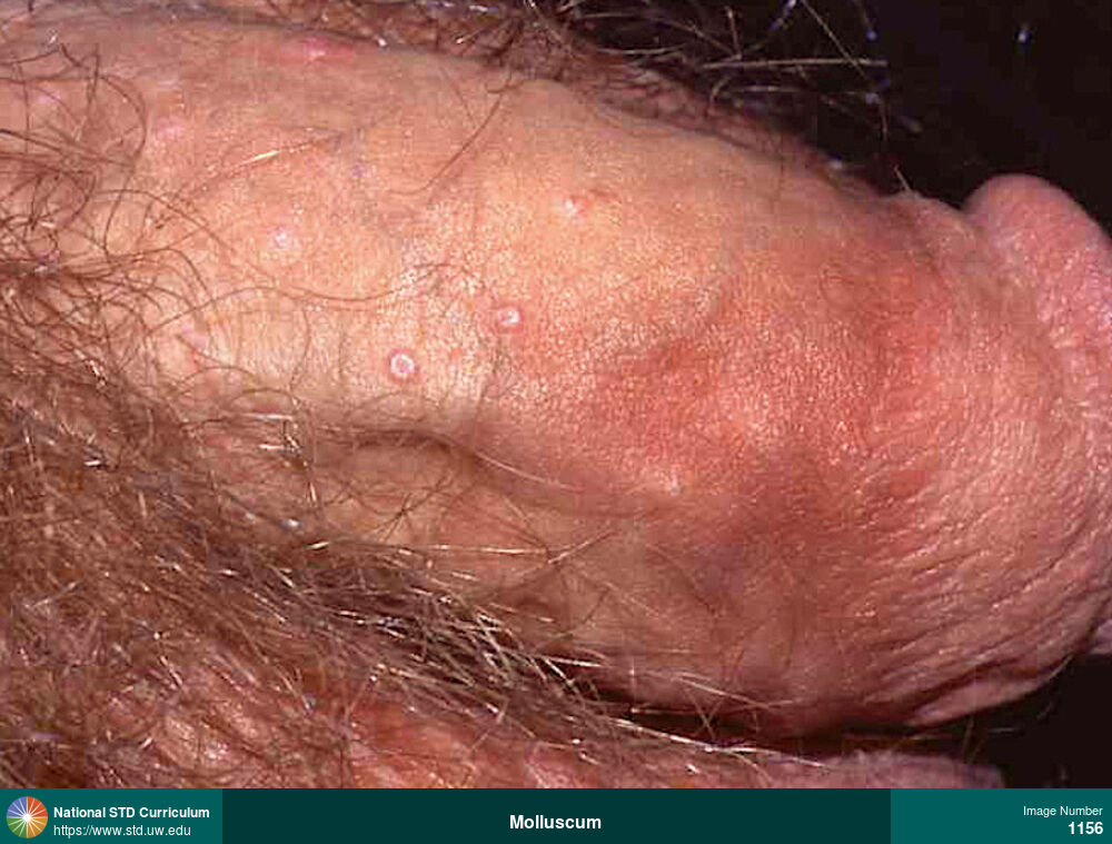
Molluscum
Molluscum contagiosum papular lesion of the penile shaft in a man with HIV. These lesions have the characteristic dome shape (with central umbilication).
Photo: Papule / Papules, Umbilicated, Light skin tone, Penis, Non-Painful
Courtesy of David H. Spach, MD
Papule / Papules, Umbilicated Light skin tone, Penis
1156
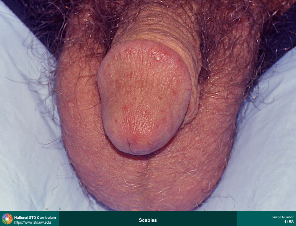
Scabies
Genital scabies manifesting as pruritic, erythematous lesions on the glans of the penis.
Photo: Erythema, Papule / Papules, Rash, Light skin tone, Penis, Itch, Non-Painful
Courtesy of David H. Spach, MD
Erythema, Papule / Papules, Rash Light skin tone, Penis
1158
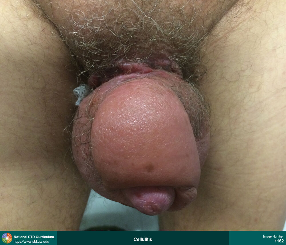
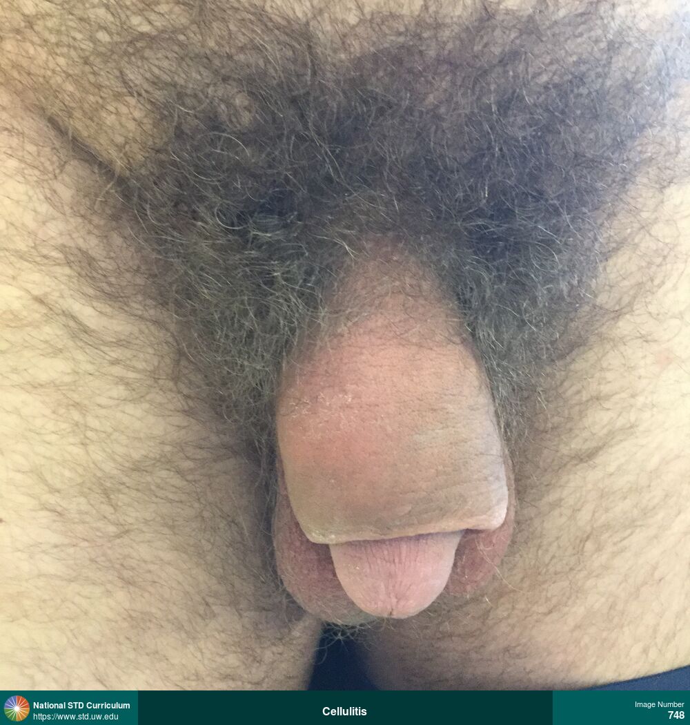
Cellulitis
Cellulitis and severe edema of penis, with necrotic tissue at the base of penis. The tissue necrosis at base of penis resulted from a retained metal cock ring; this photograph is taken after the cock ring had been cut and removed.
Photo: Edema / Swelling, Erythema, Light skin tone, Penis, Scrotum
Courtesy of David H. Spach, MD
Edema / Swelling, Erythema Light skin tone, Penis, Scrotum
1162
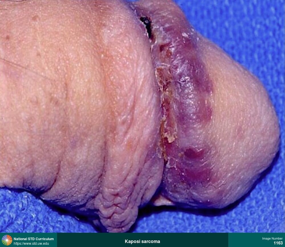
Kaposi sarcoma
Kaposi’s sarcoma in a man with HIV and AIDS. The Kaposi’s sarcoma manifested as purple-red nodular circumferential lesions around the corona of the penis.
Photo: Lesion, Plaque, Light skin tone, Penis, Non-Itchy, Non-Painful
Courtesy of David H. Spach, MD
Lesion, Plaque Light skin tone, Penis
1163
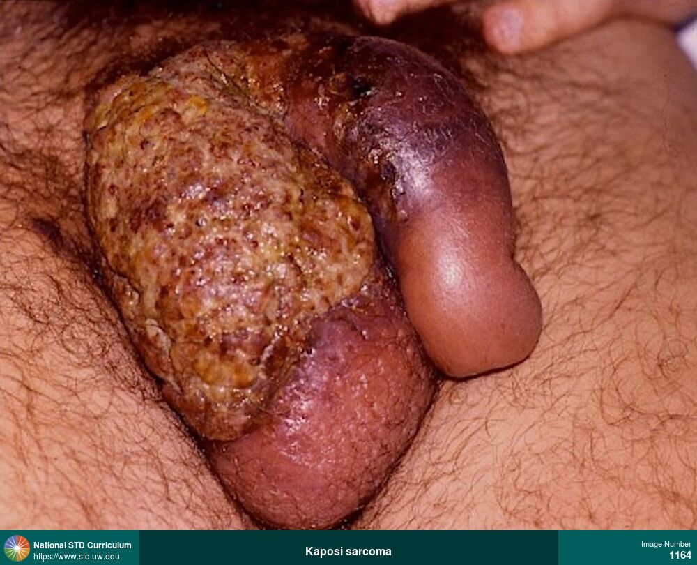
Kaposi sarcoma
Kaposi’s sarcoma of the penis and scrotum in a patient with AIDS. The Kaposi’s sarcoma has infiltrated into the deeper genital tissues, causing marked scrotal and penile edema. In addition, there is significant destruction of the skin tissue overlying the scrotum and the base of the penis.
Photo: Edema / Swelling, Lesion, Mass, Plaque, Penis, Scrotum
Courtesy of David H. Spach, MD
Edema / Swelling, Lesion, Mass, Plaque Penis, Scrotum
1164


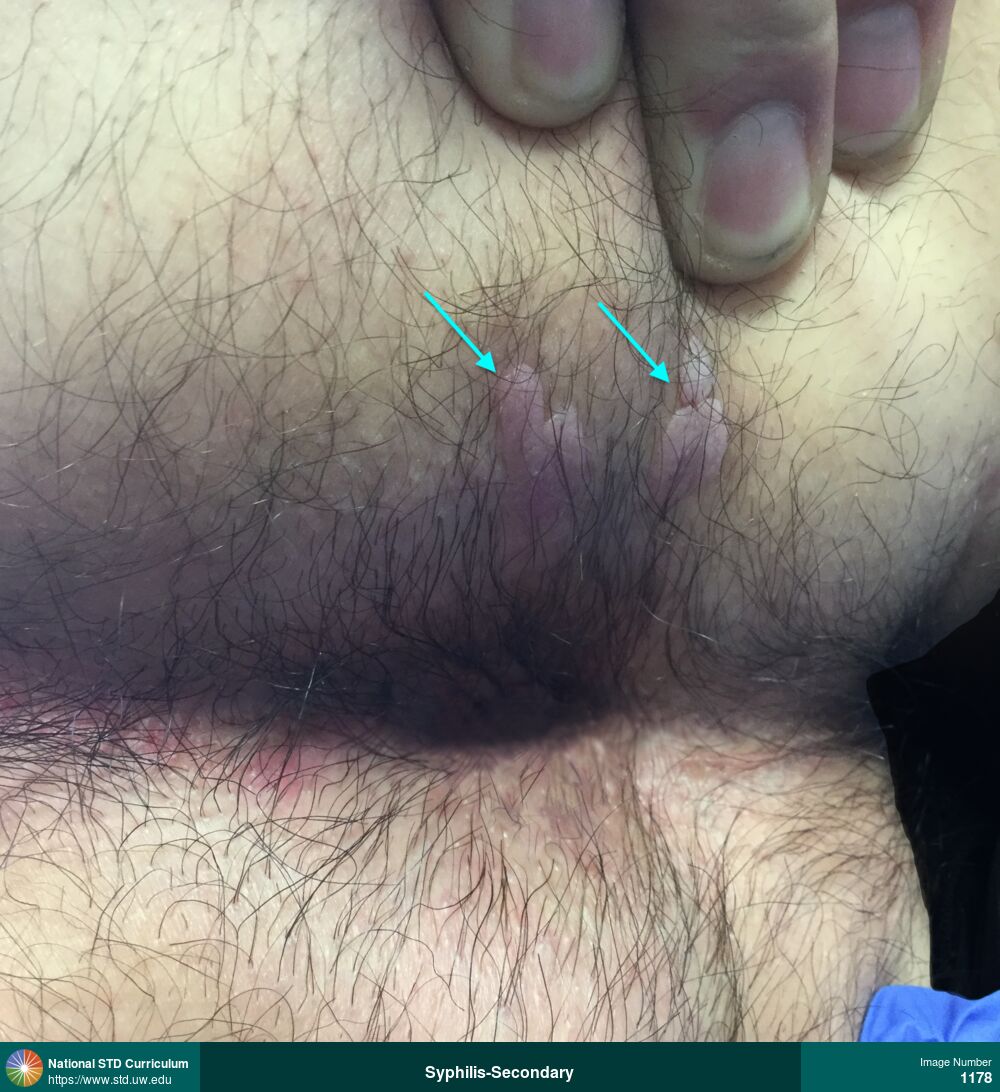
Syphilis-Secondary
Condylomata lata in man with secondary syphilis. He presented with moist, raised warty papules, most prominent in the left-sided perianal region (blue arrows). Darkfield microscopy showed abundant spirochetes.
Photo: Papule / Papules, Plaque, Anal / Perianal, Light skin tone, Tonsil, Non-Painful
Courtesy of Negusse Ocbamichael, PA
Papule / Papules, Plaque Anal / Perianal, Light skin tone, Tonsil
1178
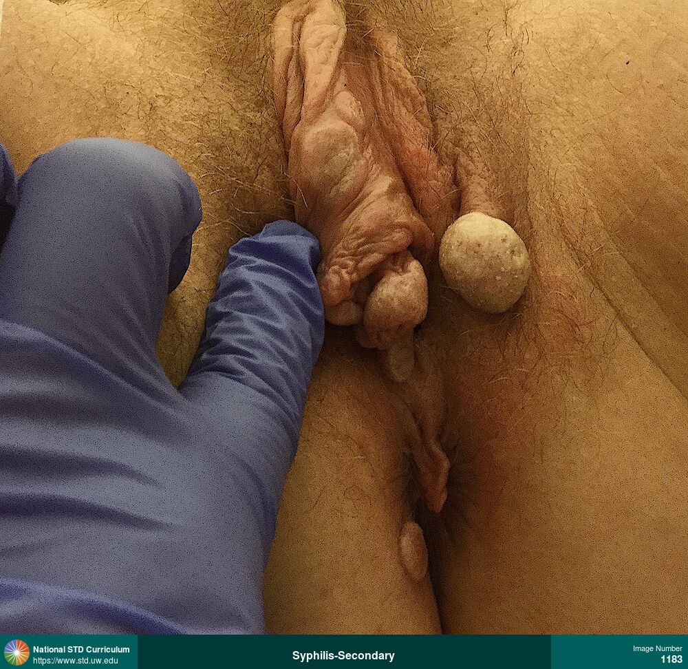
Syphilis-Secondary
Vulvar condylomata lata in a woman with secondary syphilis. This photograph shows multiple vulvar and perianal papular verrucous-appearing lesions and two large nodular vulvar lesions. Abundant spirochetes were observed on microscopy.
Photo: Papule / Papules, Plaque, Verrucous, Anal / Perianal, Light skin tone, Vulva
Courtesy of Negusse Ocbamichael, PA
Papule / Papules, Plaque, Verrucous Anal / Perianal, Light skin tone, Vulva
1183
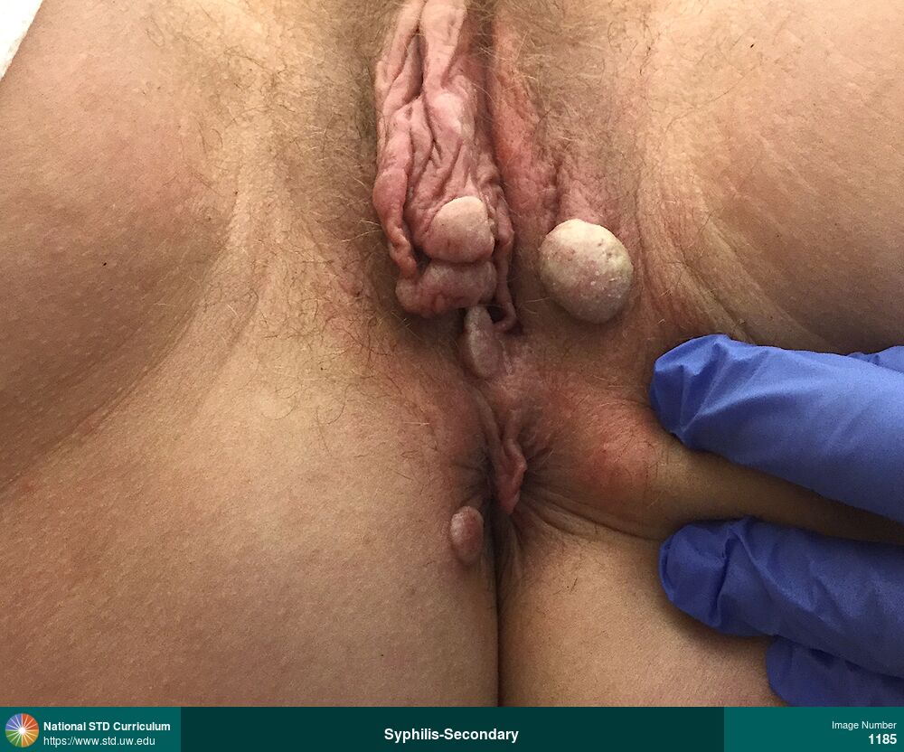
Syphilis-Secondary
Vulvar condylomata lata in a woman with secondary syphilis. This photograph shows multiple vulvar and perianal papular verrucous-appearing lesions and two large nodular vulvar lesions. Abundant spirochetes were observed on microscopy.
Photo: Papule / Papules, Plaque, Verrucous, Anal / Perianal, Light skin tone, Vulva
Courtesy of Negusse Ocbamichael, PA
Papule / Papules, Plaque, Verrucous Anal / Perianal, Light skin tone, Vulva
1185
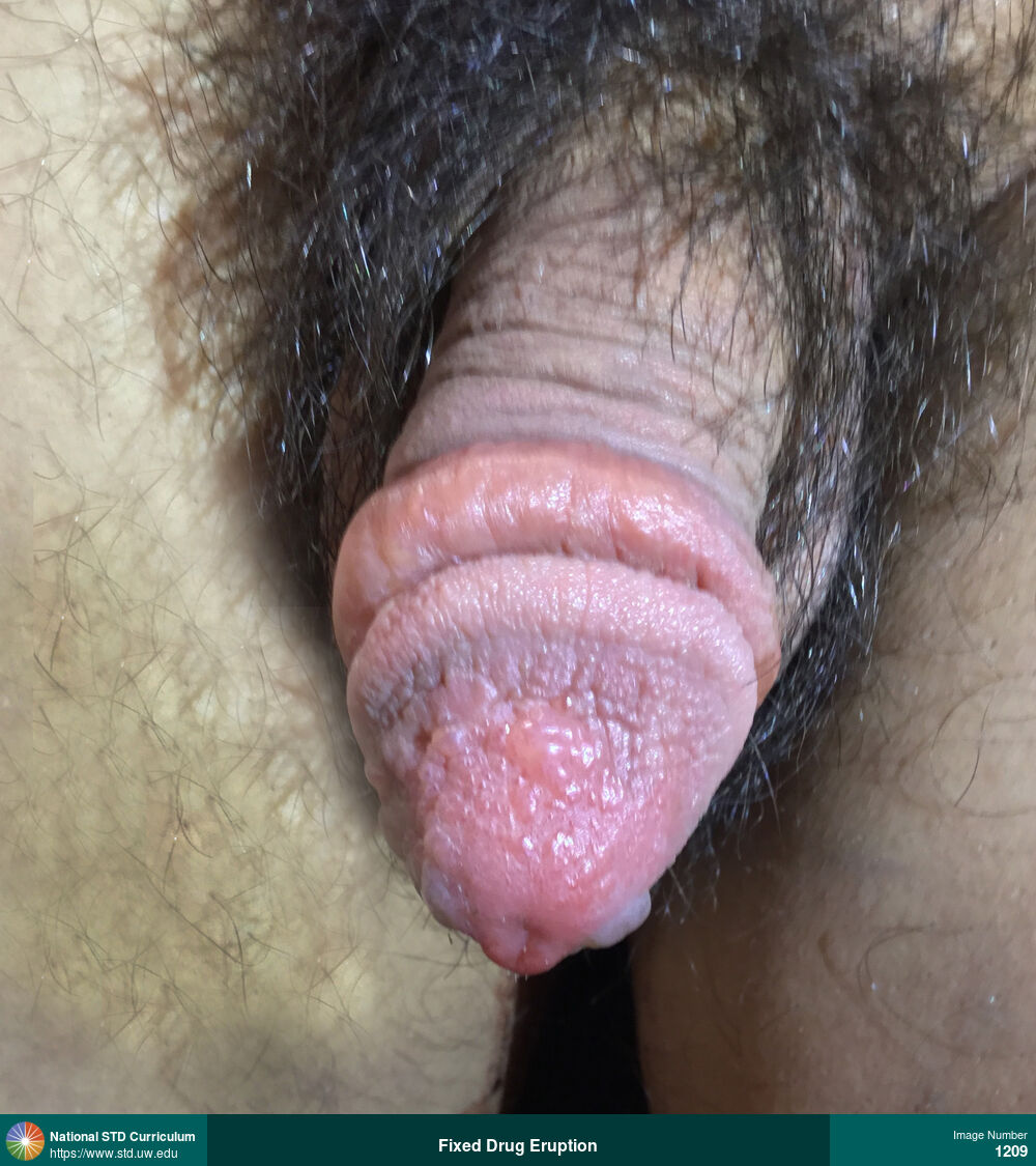
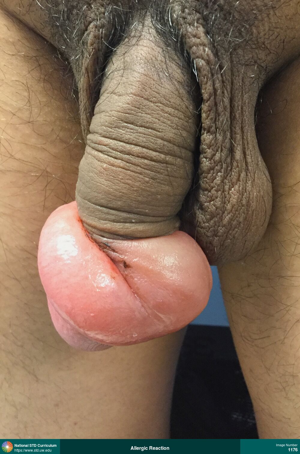
Fixed Drug Eruption
Fixed-drug eruption on glans of penis after taking trimethoprim-sulfamethoxazole. The eruption manifested as severe edema on the shaft of the penis below the glans and vesiculo-bullous lesions on the glans.
Photo: Bulla / Bullae, Edema / Swelling, Vesicle / Vesicles, Penis
Courtesy of Negusse Ocbamichael, PA
Bulla / Bullae, Edema / Swelling, Vesicle / Vesicles Penis
1209
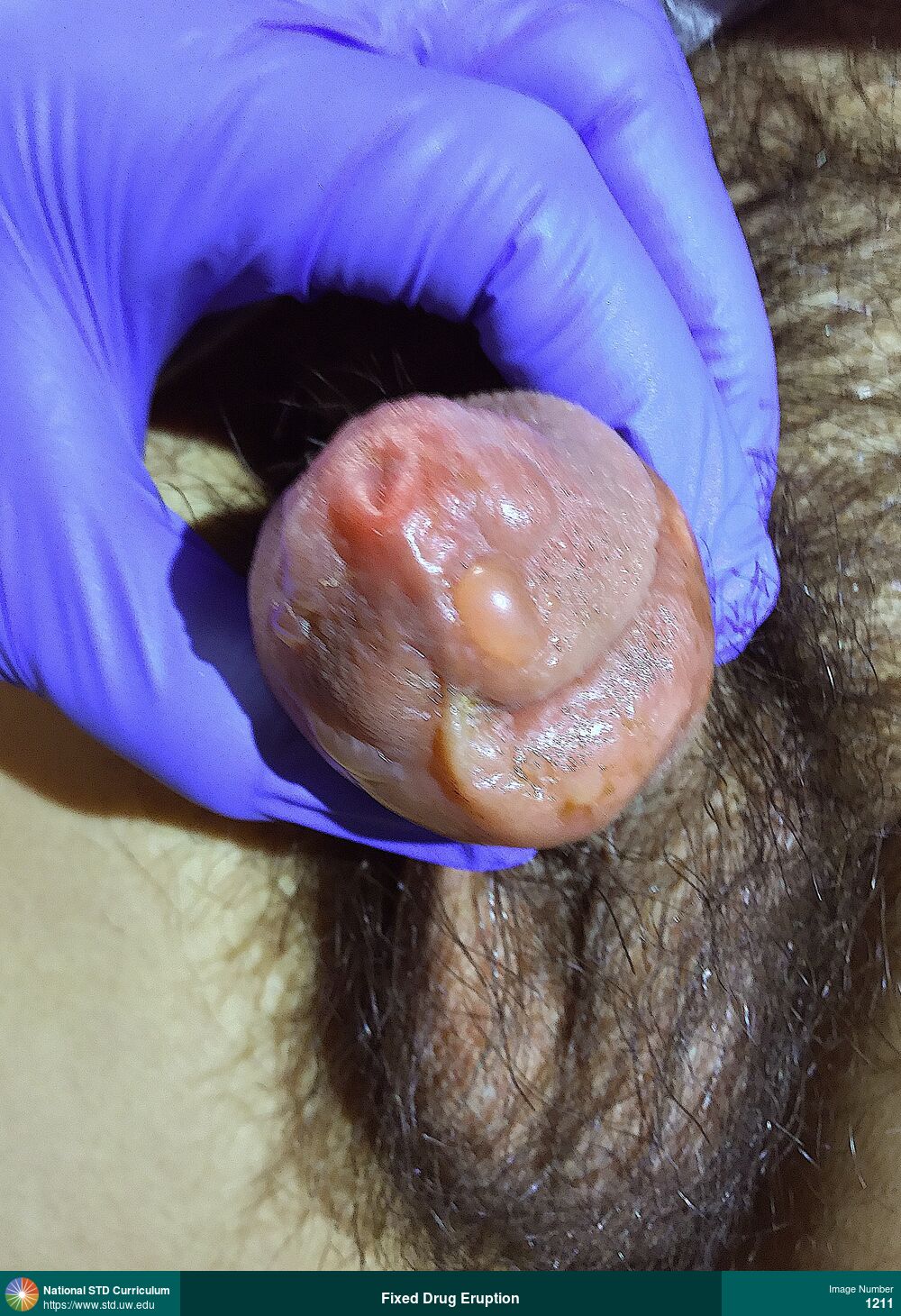
Fixed Drug Eruption
Fixed-drug eruption on glans of penis after taking trimethoprim-sulfamethoxazole. The eruption manifested as severe edema on the shaft of the penis below the glans and vesiculo-bullous lesions on the glans.
Photo: Bulla / Bullae, Edema / Swelling, Penis
Courtesy of Negusse Ocbamichael, PA
Bulla / Bullae, Edema / Swelling Penis
1211
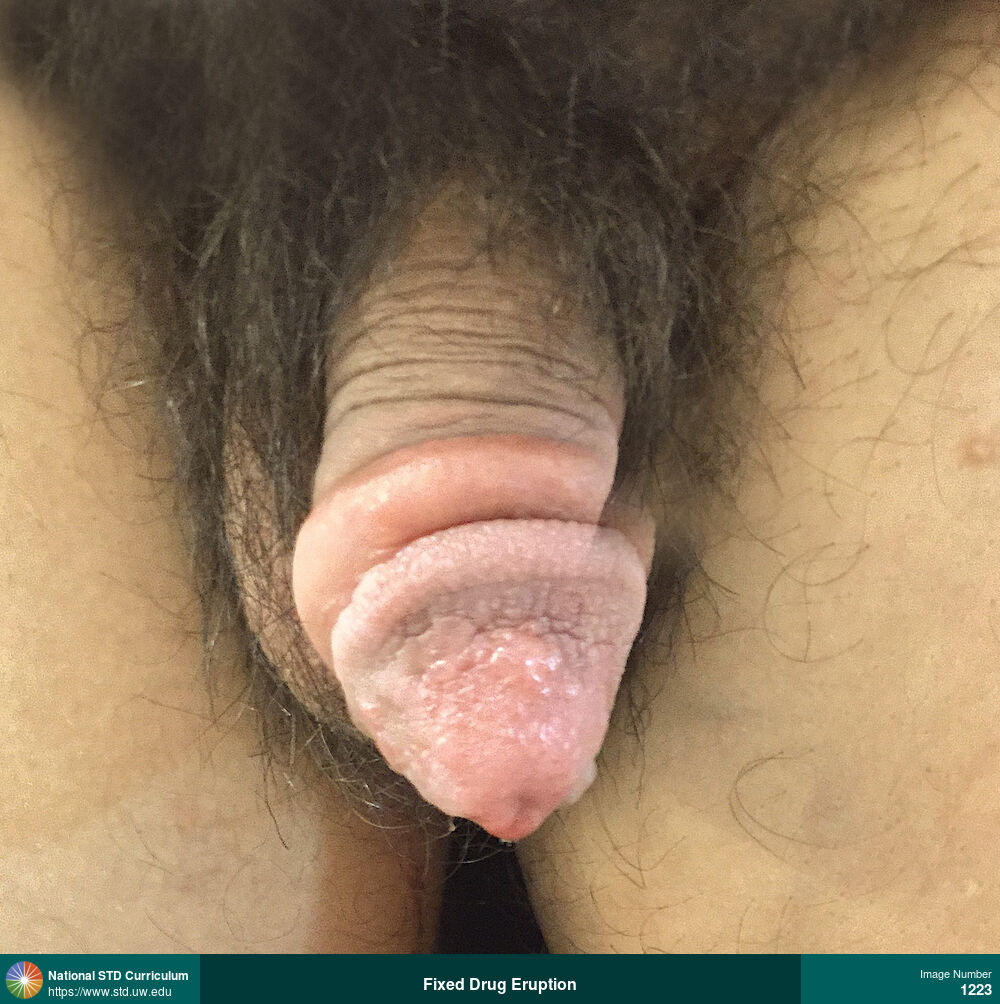
Fixed Drug Eruption
Fixed-drug eruption on glans of penis after taking trimethoprim-sulfamethoxazole. The eruption manifested as severe edema on the shaft of the penis below the glans and vesiculo-bullous lesions on the glans.
Photo: Bulla / Bullae, Edema / Swelling, Vesicle / Vesicles, Penis
Courtesy of Negusse Ocbamichael, PA
Bulla / Bullae, Edema / Swelling, Vesicle / Vesicles Penis
1223
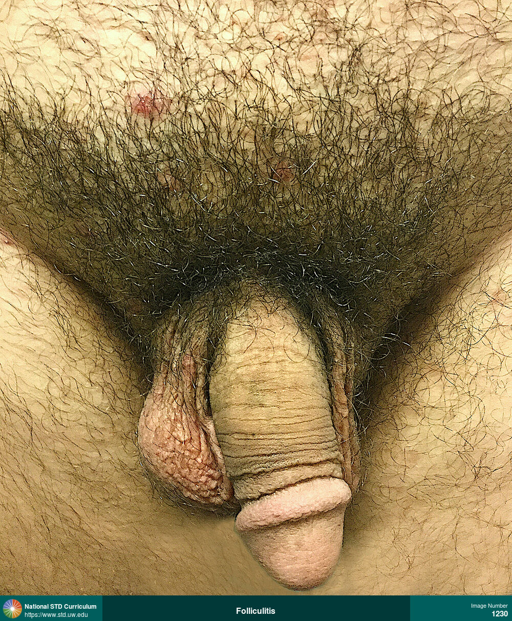
Folliculitis
Photo: Papule / Papules, Light skin tone, Suprapubic (Hypogastrium)
Courtesy of Negusse Ocbamichael, PA
Papule / Papules Light skin tone, Suprapubic (Hypogastrium)
1230
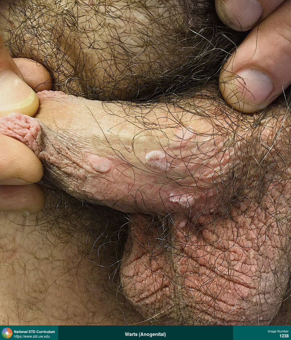
Warts (Anogenital)
Multiple warts on the shaft of the penis.
Photo: Papule / Papules, Verrucous, Light skin tone, Penis, Non-Painful
Courtesy of Negusse Ocbamichael, PA
Papule / Papules, Verrucous Light skin tone, Penis
1238
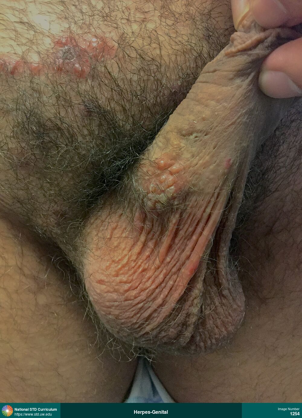
Herpes-Genital
Photo: Erythema, Vesicle / Vesicles, Light skin tone, Penis, Suprapubic (Hypogastrium)
Courtesy of Negusse Ocbamichael, PA
Erythema, Vesicle / Vesicles Light skin tone, Penis, Suprapubic (Hypogastrium)
1254
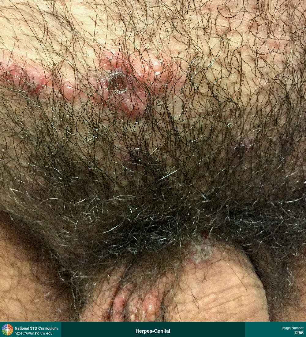
Herpes-Genital
Photo: Erythema, Vesicle / Vesicles, Light skin tone, Penis, Suprapubic (Hypogastrium)
Courtesy of Negusse Ocbamichael, PA
Erythema, Vesicle / Vesicles Light skin tone, Penis, Suprapubic (Hypogastrium)
1255

Herpes-Genital
Photo: Erythema, Vesicle / Vesicles, Light skin tone, Penis, Scrotum
Courtesy of Negusse Ocbamichael, PA
Erythema, Vesicle / Vesicles Light skin tone, Penis, Scrotum
1256
Herpes-Genital
Photo: Erythema, Vesicle / Vesicles, Light skin tone, Penis, Scrotum, Suprapubic (Hypogastrium)
Courtesy of Negusse Ocbamichael, PA
Erythema, Vesicle / Vesicles Light skin tone, Penis, Scrotum, Suprapubic (Hypogastrium)
1258

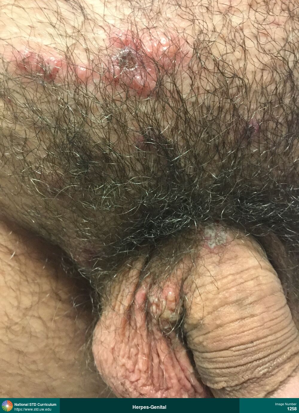
Herpes-Genital
Photo: Edema / Swelling, Scab, Ulcer / Ulcers, Light skin tone, Penis
Courtesy of Negusse Ocbamichael, PA
Edema / Swelling, Scab, Ulcer / Ulcers Light skin tone, Penis
1259

Herpes-Genital
Photo: Edema / Swelling, Ulcer / Ulcers, Light skin tone, Penis
Courtesy of Negusse Ocbamichael, PA
Edema / Swelling, Ulcer / Ulcers Light skin tone, Penis
1261
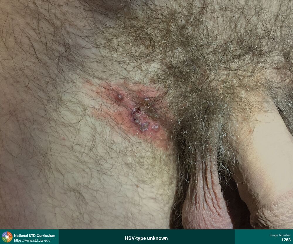
Herpes-Body
Photo: Erythema, Vesicle / Vesicles, Groin/Inguinal, Light skin tone, Rash
Courtesy of Negusse Ocbamichael, PA
Erythema, Vesicle / Vesicles Groin/Inguinal, Light skin tone
1263
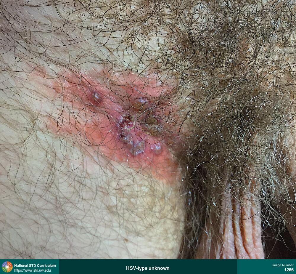
Herpes-Body
Photo: Erythema, Scab, Vesicle / Vesicles, Groin/Inguinal, Light skin tone, Rash
Courtesy of Negusse Ocbamichael, PA
Erythema, Scab, Vesicle / Vesicles Groin/Inguinal, Light skin tone
1266
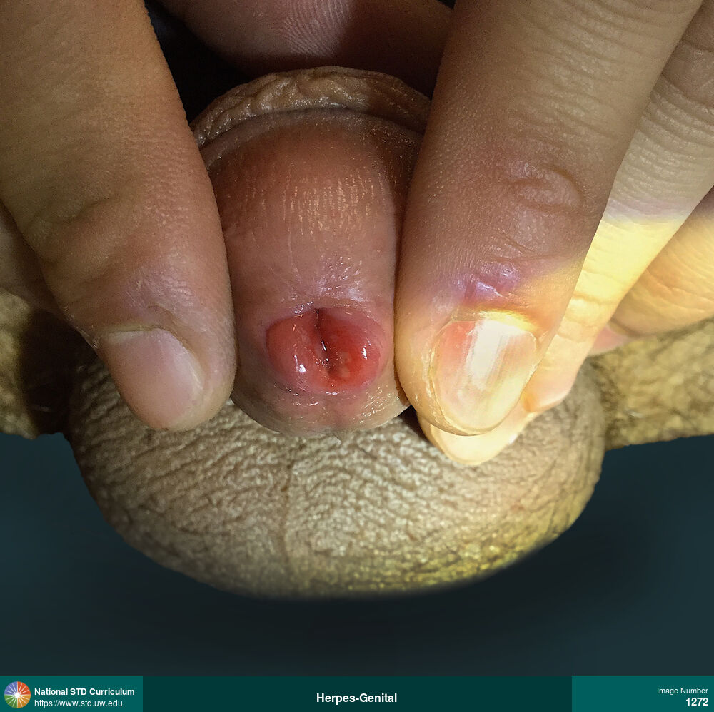
Herpes-Digital (Whitlow), Herpes-Genital
Photo: Ulcer / Ulcers, Light skin tone, Penis, Dysuria
Courtesy of Negusse Ocbamichael, PA
Ulcer / Ulcers Light skin tone, Penis
1272
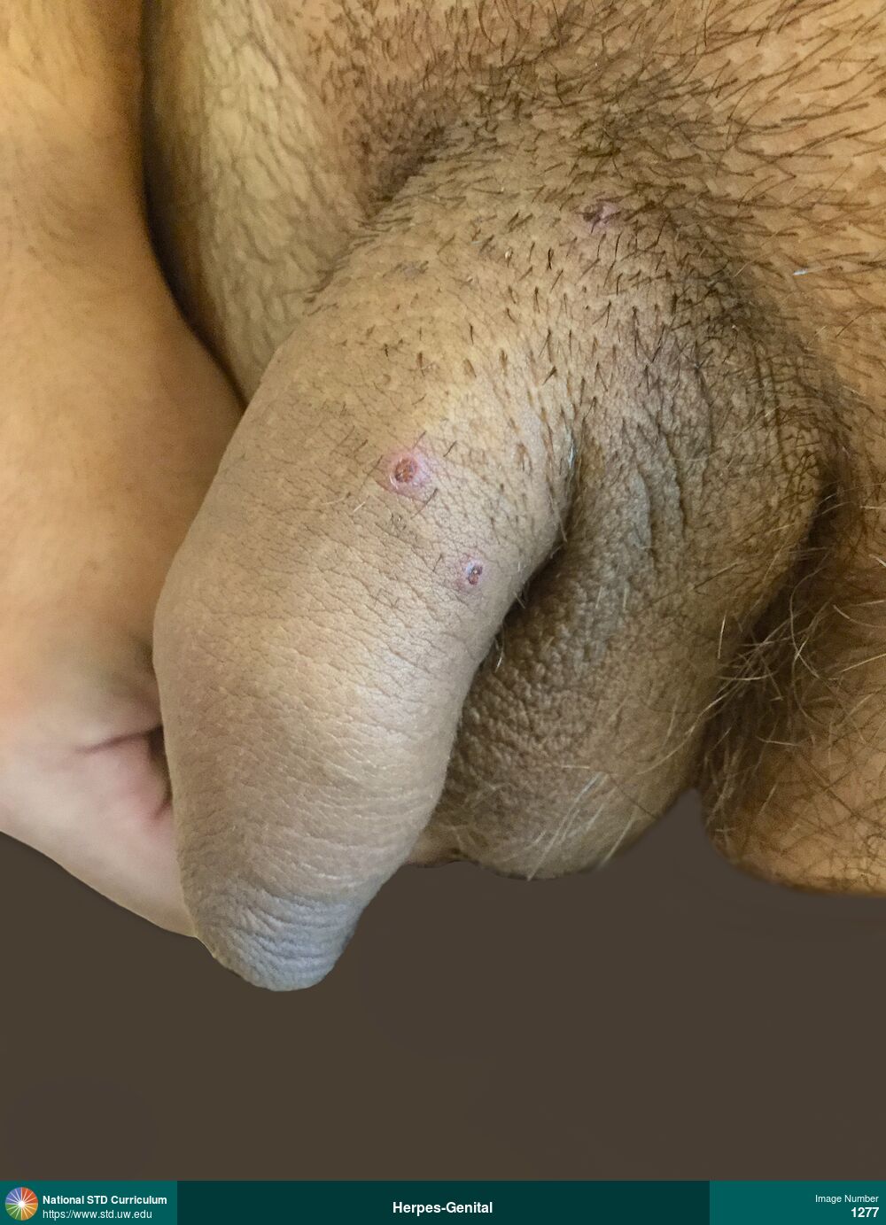
Herpes-Digital (Whitlow), Herpes-Genital
Photo: Ulcer / Ulcers, Light skin tone, Penis, Dysuria, Painful
Courtesy of Negusse Ocbamichael, PA
Ulcer / Ulcers Light skin tone, Penis
1277
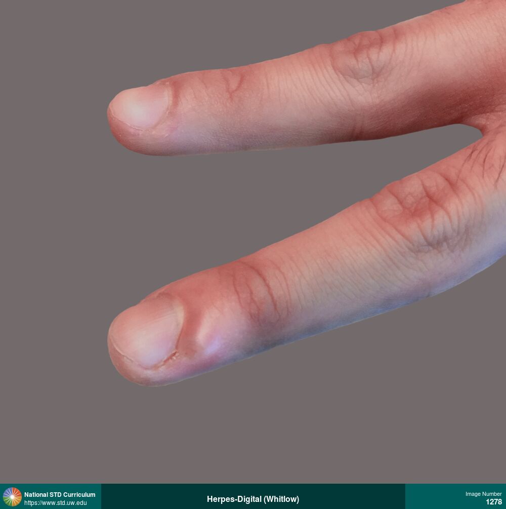
Herpes-Digital (Whitlow), Herpes-Genital
Photo: Edema / Swelling, Vesicle / Vesicles, Hand (Left), Light skin tone, Non-Itchy, Painful
Courtesy of Negusse Ocbamichael, PA
Edema / Swelling, Vesicle / Vesicles Hand (Left), Light skin tone
1278
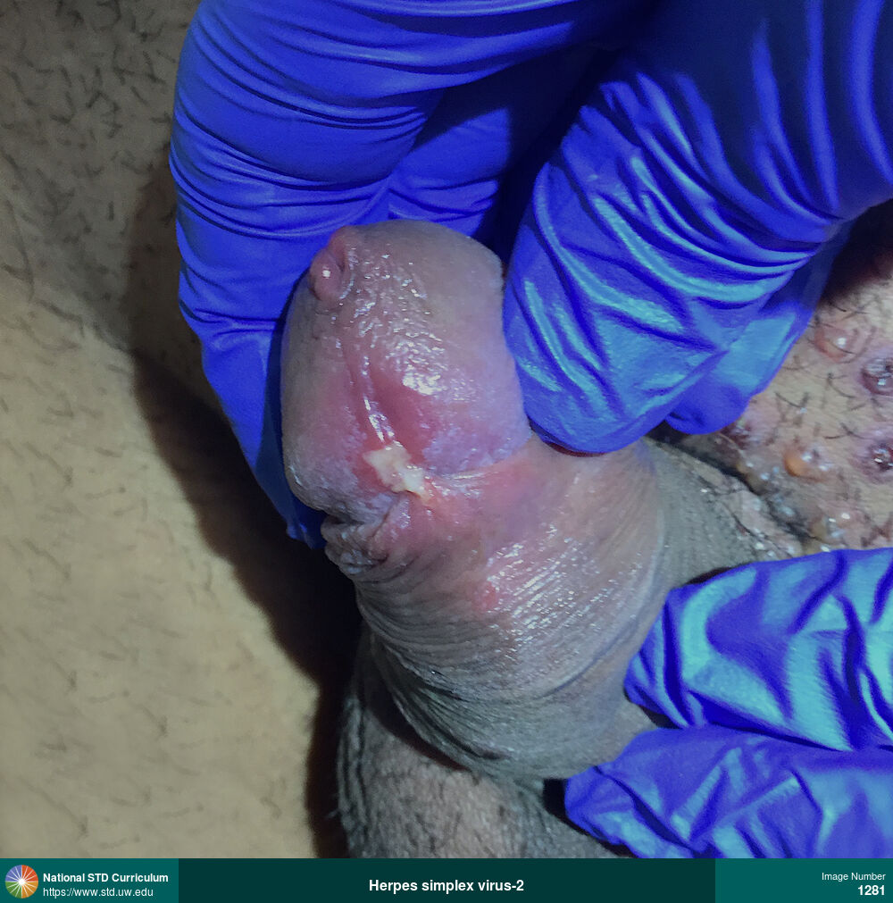
Herpes-Genital
Photo: Erythema, Ulcer / Ulcers, Vesicle / Vesicles, Penis, Painful
Courtesy of Negusse Ocbamichael, PA
Erythema, Ulcer / Ulcers, Vesicle / Vesicles Penis
1281
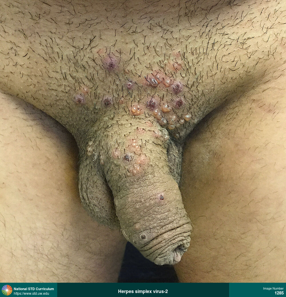
Herpes-Genital
Photo: Erythema, Ulcer / Ulcers, Vesicle / Vesicles, Light skin tone, Penis, Suprapubic (Hypogastrium), Painful
Courtesy of Negusse Ocbamichael, PA
Erythema, Ulcer / Ulcers, Vesicle / Vesicles Light skin tone, Penis, Suprapubic (Hypogastrium)
1285
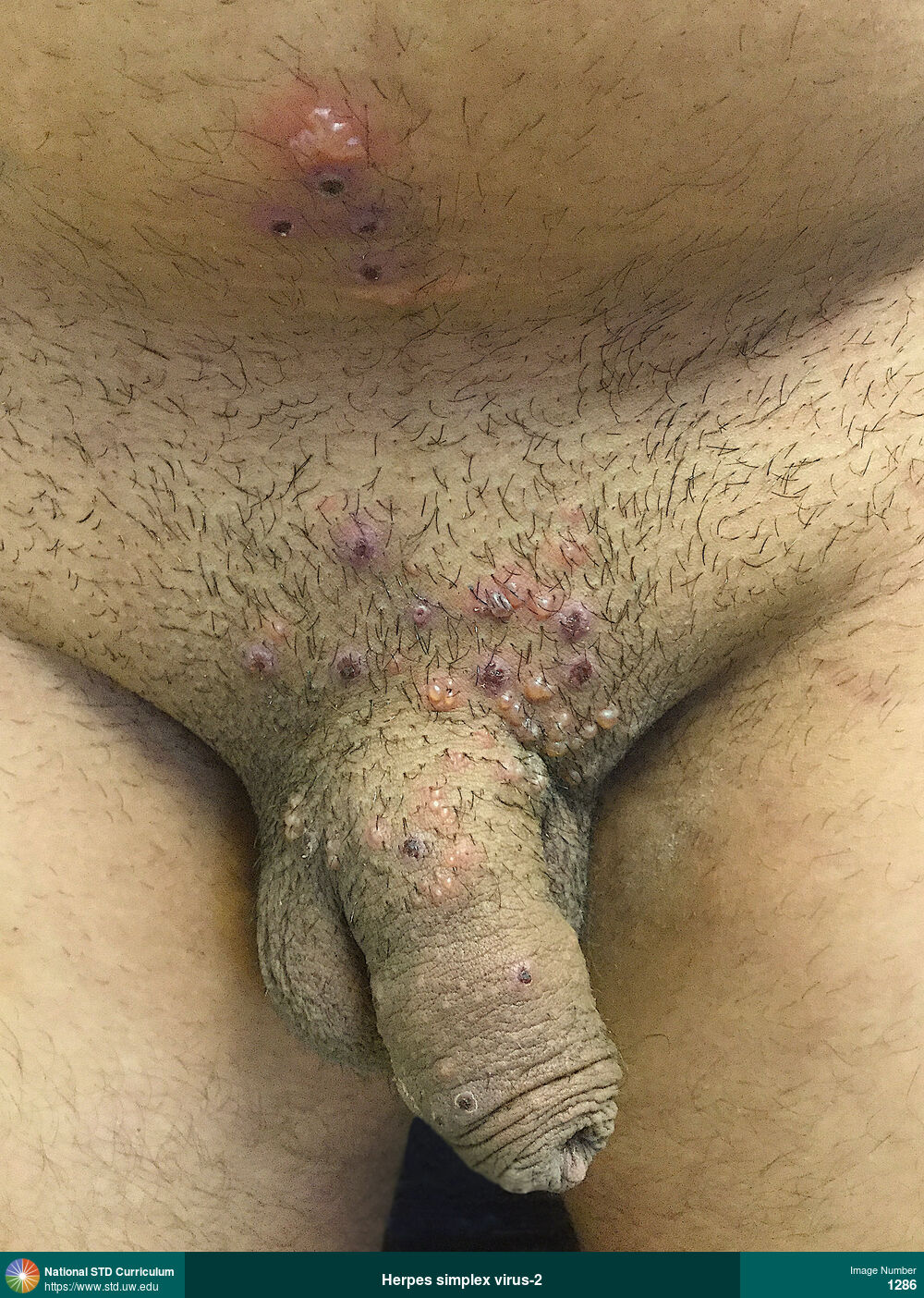
Herpes-Genital
Photo: Erythema, Ulcer / Ulcers, Vesicle / Vesicles, Light skin tone, Penis, Suprapubic (Hypogastrium), Painful
Courtesy of Negusse Ocbamichael, PA
Erythema, Ulcer / Ulcers, Vesicle / Vesicles Light skin tone, Penis, Suprapubic (Hypogastrium)
1286

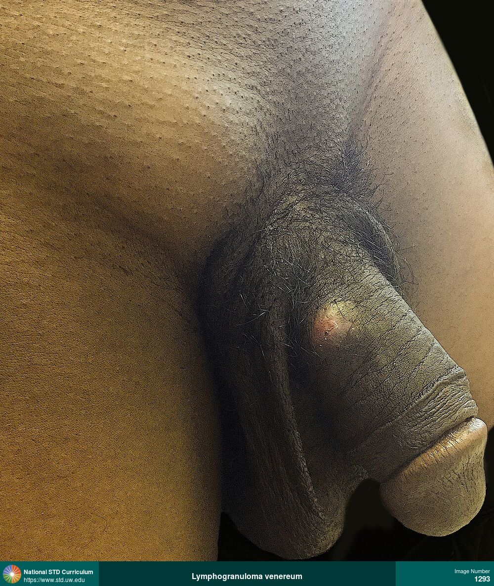
Chlamydia, Lymphogranuloma venereum
Photo: Adenopathy, Ulcer / Ulcers, Groin/Inguinal, Penis, Painful
Courtesy of Negusse Ocbamichael, PA
Adenopathy, Ulcer / Ulcers Groin/Inguinal, Penis
1293
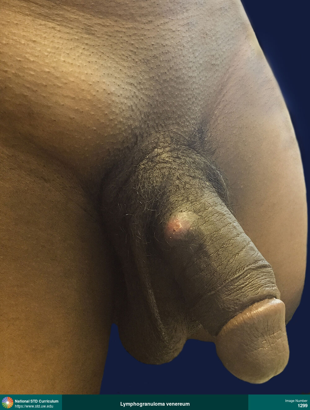
Chlamydia, Lymphogranuloma venereum
Photo: Adenopathy, Plaque, Ulcer / Ulcers, Dark skin tone, Groin/Inguinal, Penis, Painful
Courtesy of Negusse Ocbamichael, PA
Adenopathy, Plaque, Ulcer / Ulcers Dark skin tone, Groin/Inguinal, Penis
1299
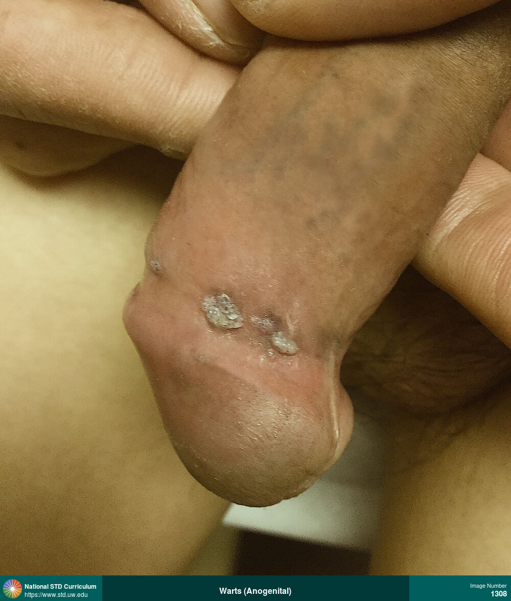
Warts (Anogenital)
A cluster of warts on the distal shaft of the penis just proximal the glans.
Photo: Papule / Papules, Verrucous, Dark skin tone, Penis, Non-Itchy, Non-Painful
Courtesy of Negusse Ocbamichael, PA
Papule / Papules, Verrucous Dark skin tone, Penis
1308
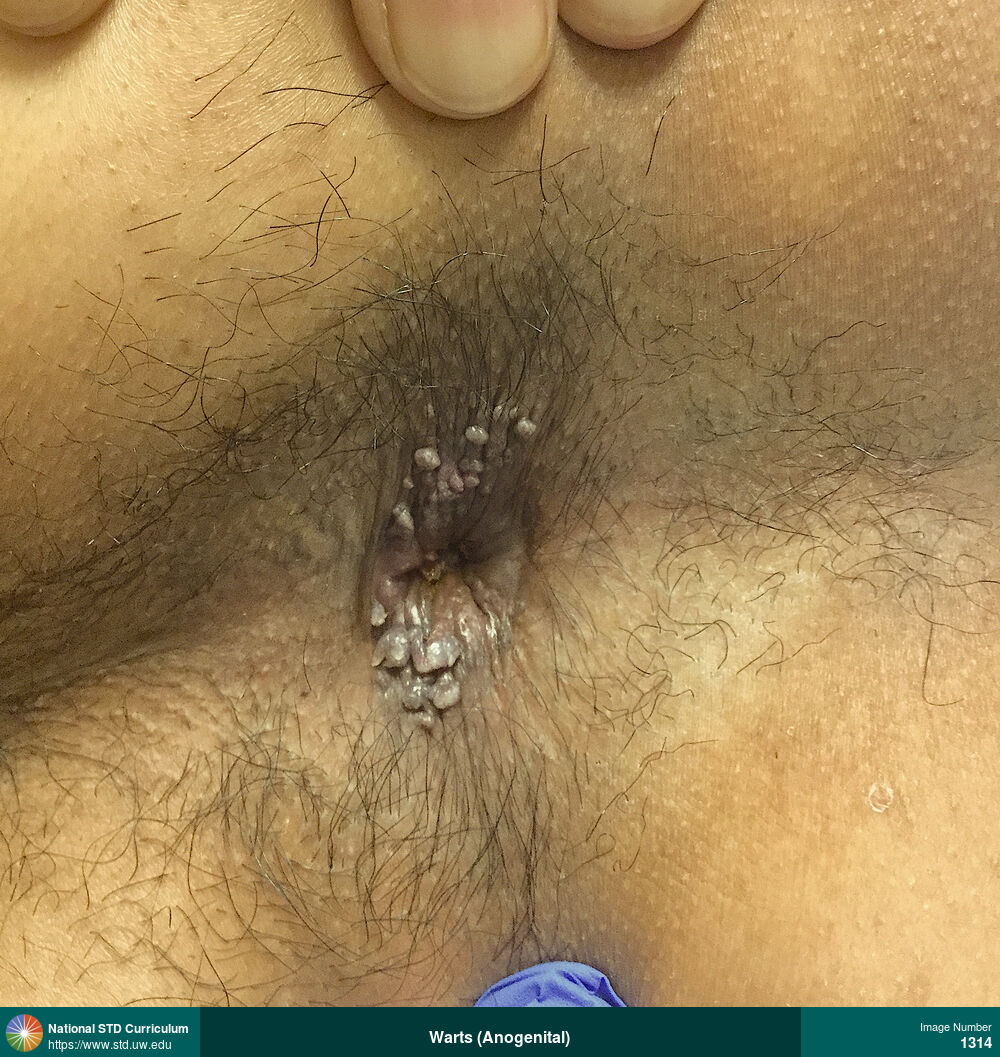
Warts (Anogenital)
Multiple perianal and intra-anal warts.
Photo: Papule / Papules, Verrucous, Anal / Intra-anal, Anal / Perianal, Dark skin tone, Non-Itchy, Non-Painful
Courtesy of Negusse Ocbamichael, PA
Papule / Papules, Verrucous Anal / Intra-anal, Anal / Perianal, Dark skin tone
1314
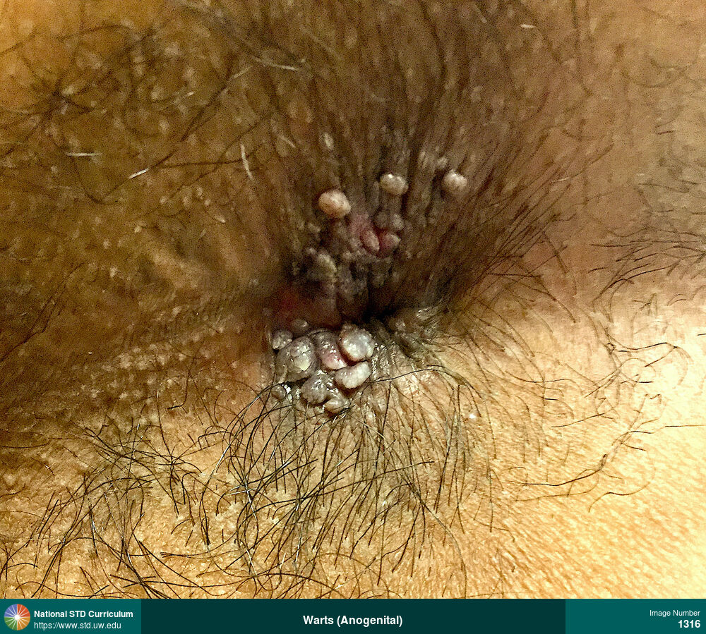
Warts (Anogenital)
Multiple perianal and intra-anal warts.
Photo: Papule / Papules, Verrucous, Anal / Perianal, Dark skin tone, Non-Itchy, Non-Painful
Courtesy of Negusse Ocbamichael, PA
Papule / Papules, Verrucous Anal / Perianal, Dark skin tone
1316
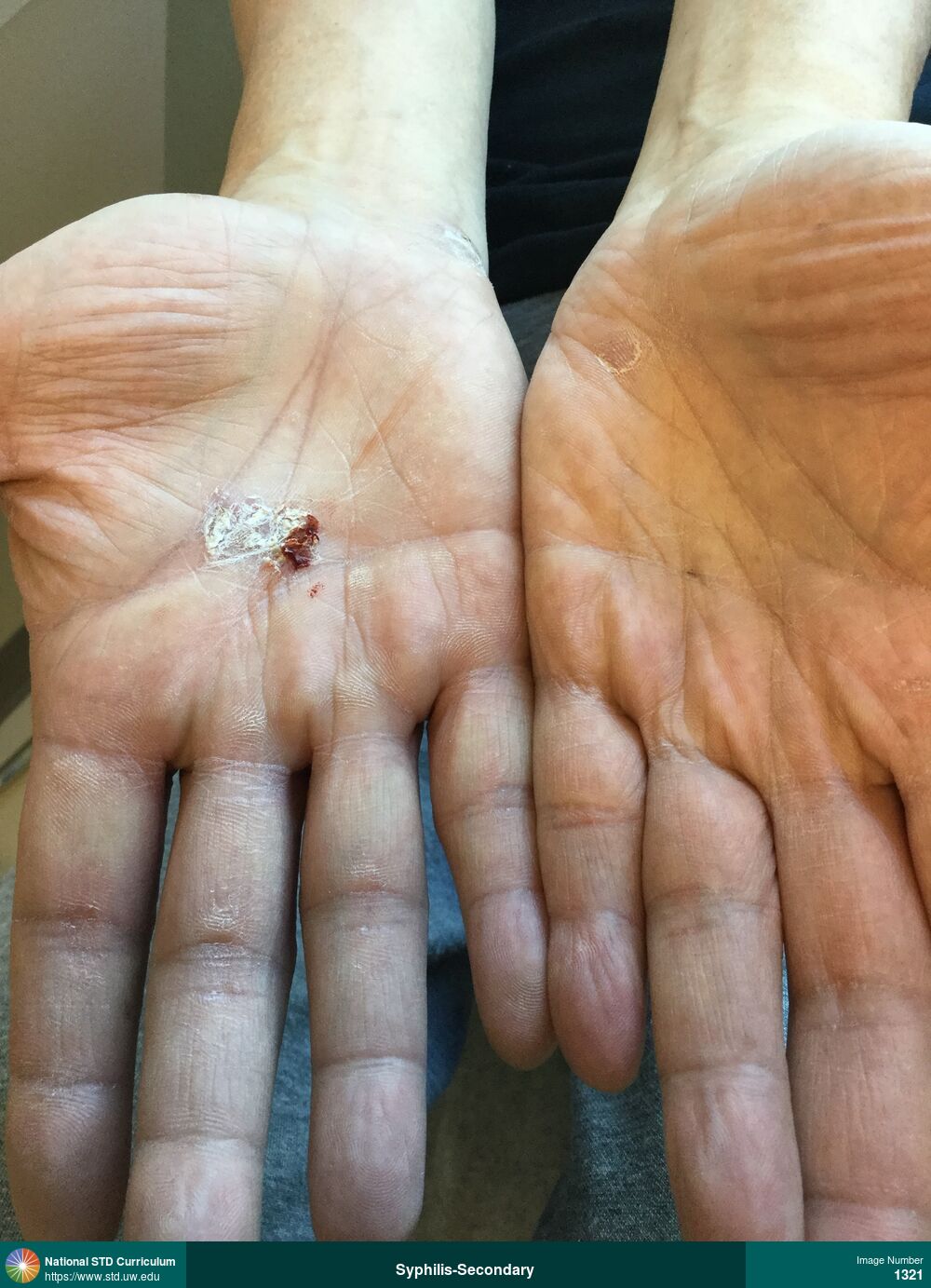
Syphilis-Secondary
Photo: Papule / Papules, Plaque, Hand (Right)
Courtesy of Negusse Ocbamichael, PA
Papule / Papules, Plaque Hand (Right)
1321
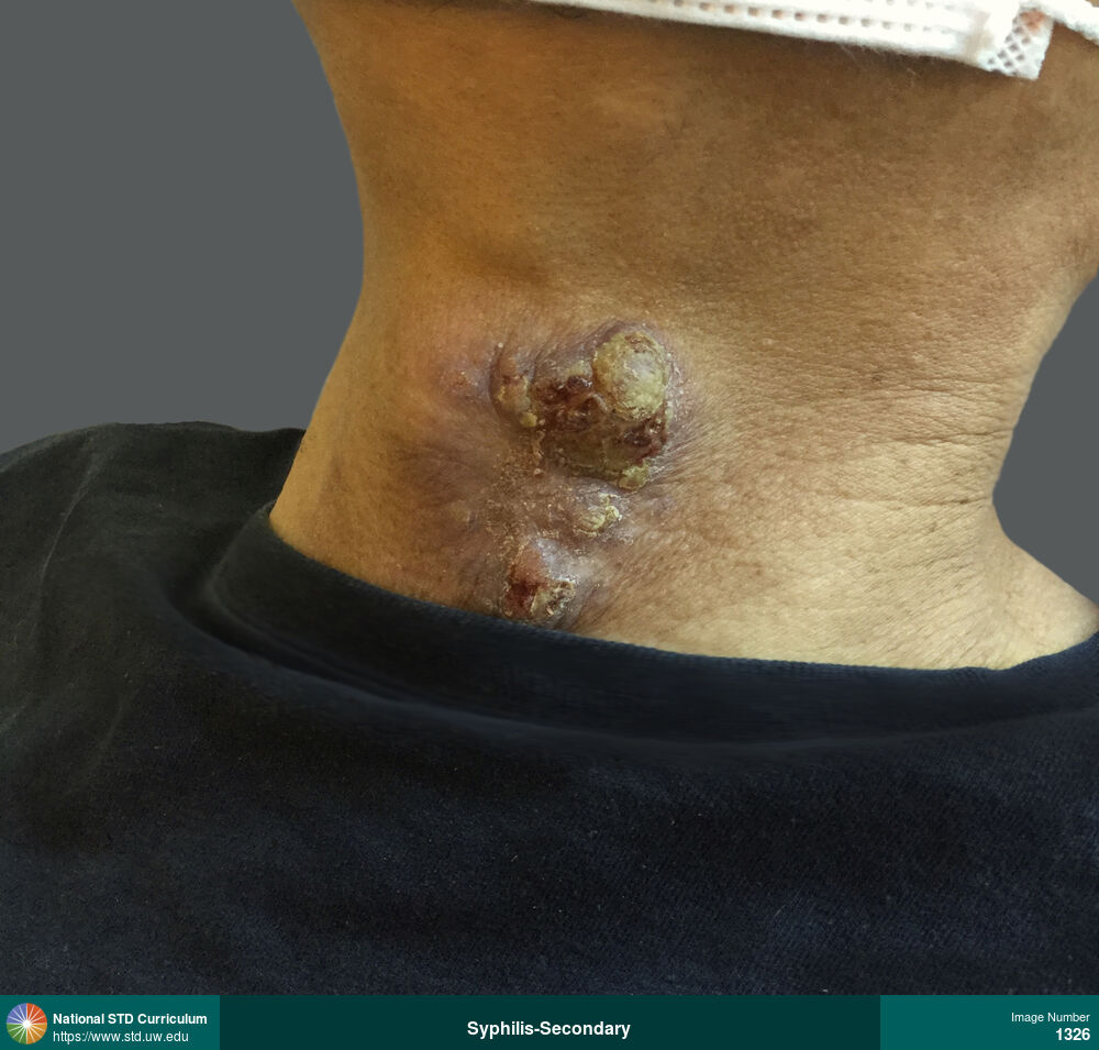
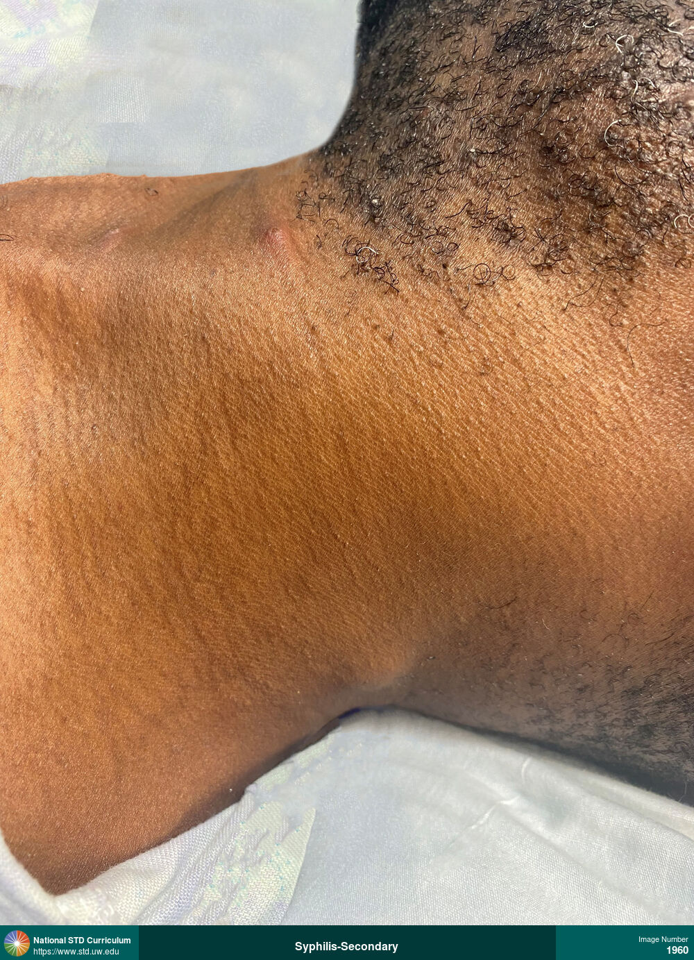
Syphilis-Secondary
Photo: Edema / Swelling, Lesion, Light skin tone, Neck
Courtesy of Negusse Ocbamichael, PA
Edema / Swelling, Lesion Light skin tone, Neck
1326
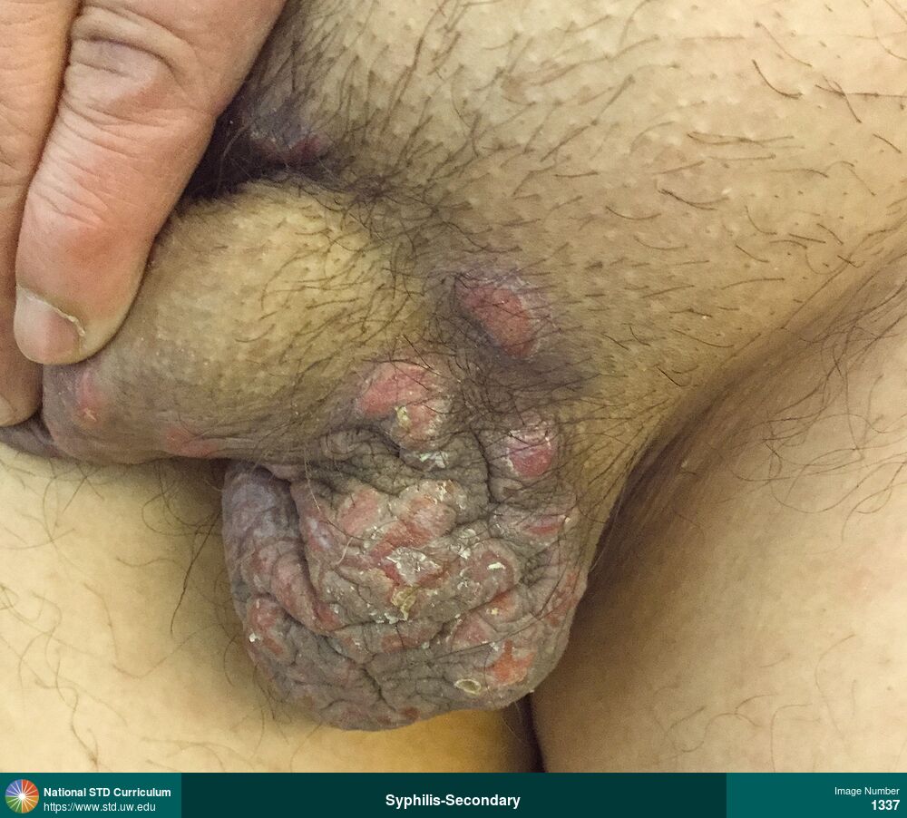
Syphilis-Secondary
Photo: Cyst, Papule / Papules, Plaque, Scrotum, Rash
Courtesy of Negusse Ocbamichael, PA
Cyst, Papule / Papules, Plaque Scrotum
1337
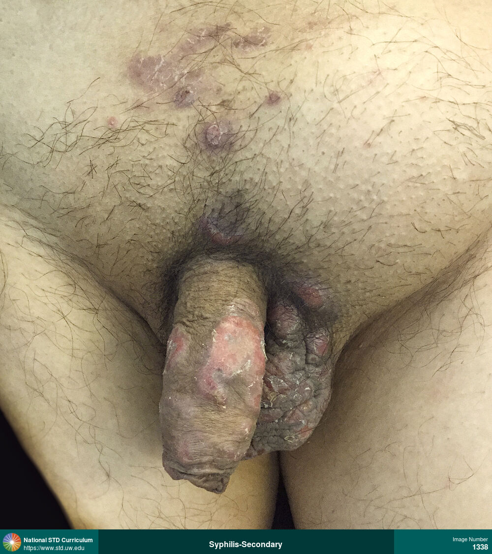
Syphilis-Secondary
Photo: Papule / Papules, Light skin tone, Penis, Scrotum, Suprapubic (Hypogastrium), Rash
Courtesy of Negusse Ocbamichael, PA
Papule / Papules Light skin tone, Penis, Scrotum, Suprapubic (Hypogastrium)
1338
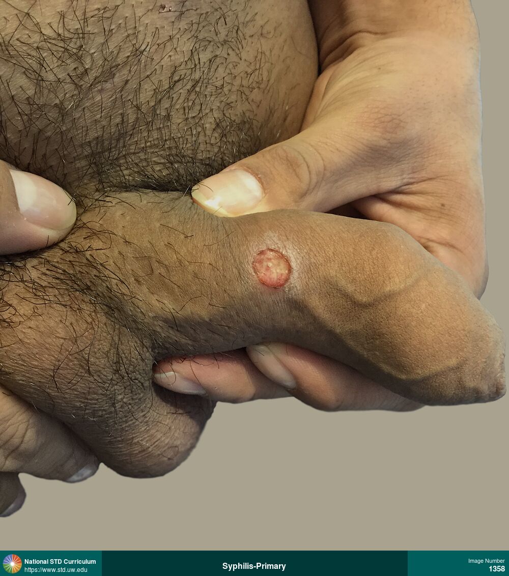
Syphilis-Primary
Photo: Ulcer / Ulcers, Penis, Non-Painful
Courtesy of Negusse Ocbamichael, PA
Ulcer / Ulcers Penis
1358
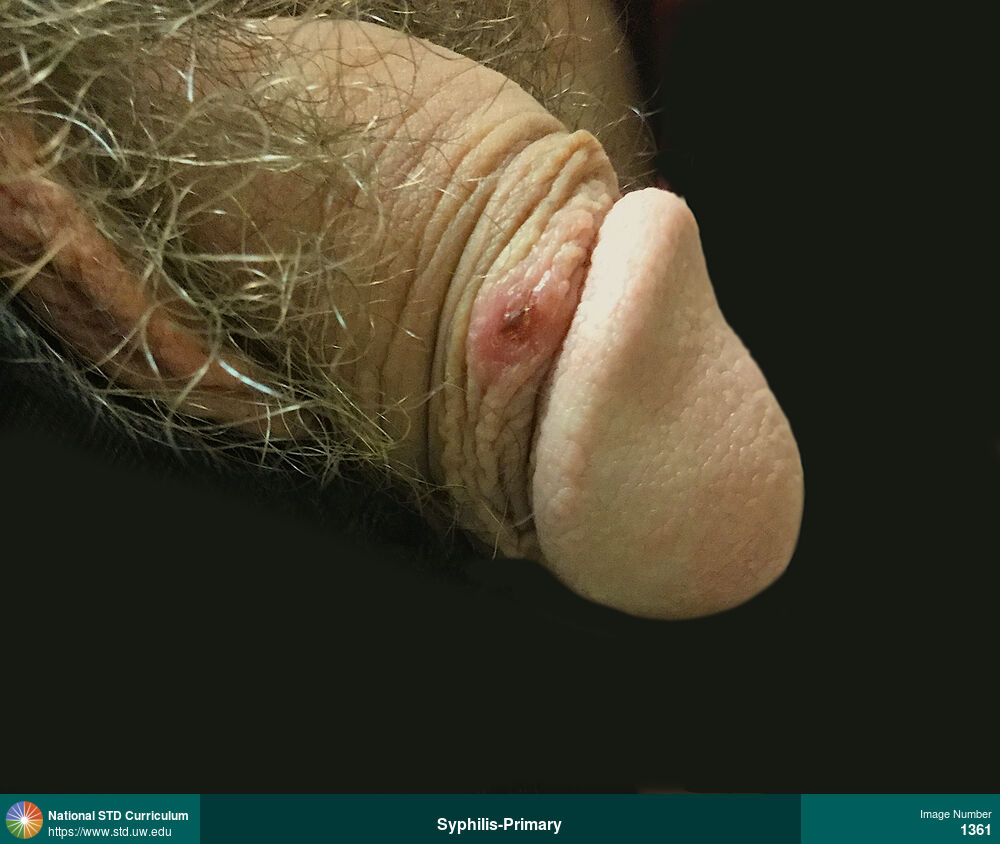
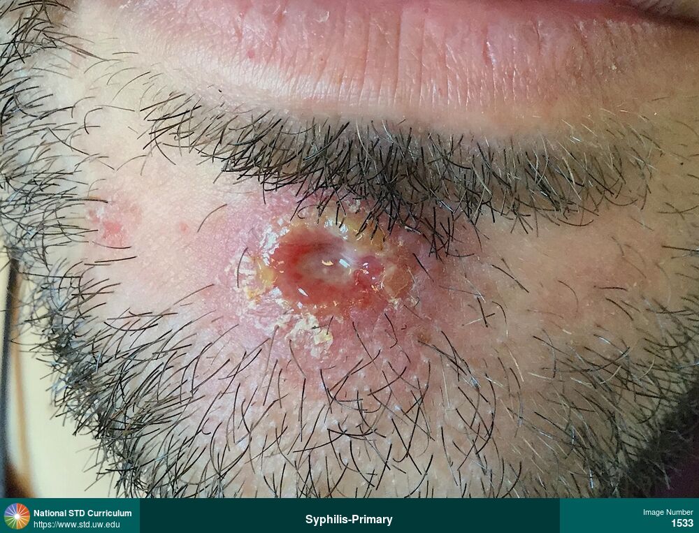
Syphilis-Primary
Photo: Erythema, Lesion, Scab, Ulcer / Ulcers, Light skin tone, Penis, Non-Painful
Courtesy of Negusse Ocbamichael, PA
Erythema, Lesion, Scab, Ulcer / Ulcers Light skin tone, Penis
1361
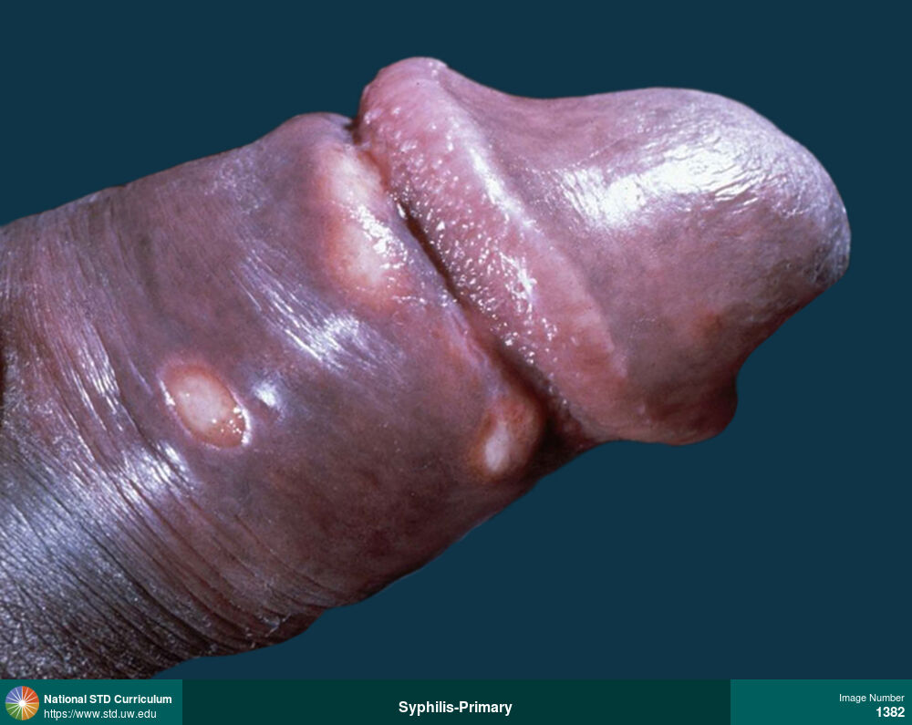
Syphilis-Primary
Photo: Scale, Ulcer / Ulcers, Dark skin tone, Penis
Courtesy of Negusse Ocbamichael, PA
Scale, Ulcer / Ulcers Dark skin tone, Penis
1382

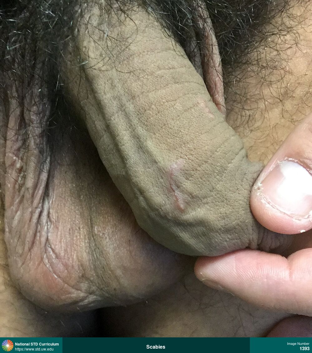
Scabies
Linear burrow on the shaft of the penis caused by scabies. The rash on the penis was intensely pruritic. This man also had papules and burrows on his hands bilaterally.
Photo: Papule / Papules, Dark skin tone, Penis, Itch
Courtesy of Negusse Ocbamichael, PA
Papule / Papules Dark skin tone, Penis
1393
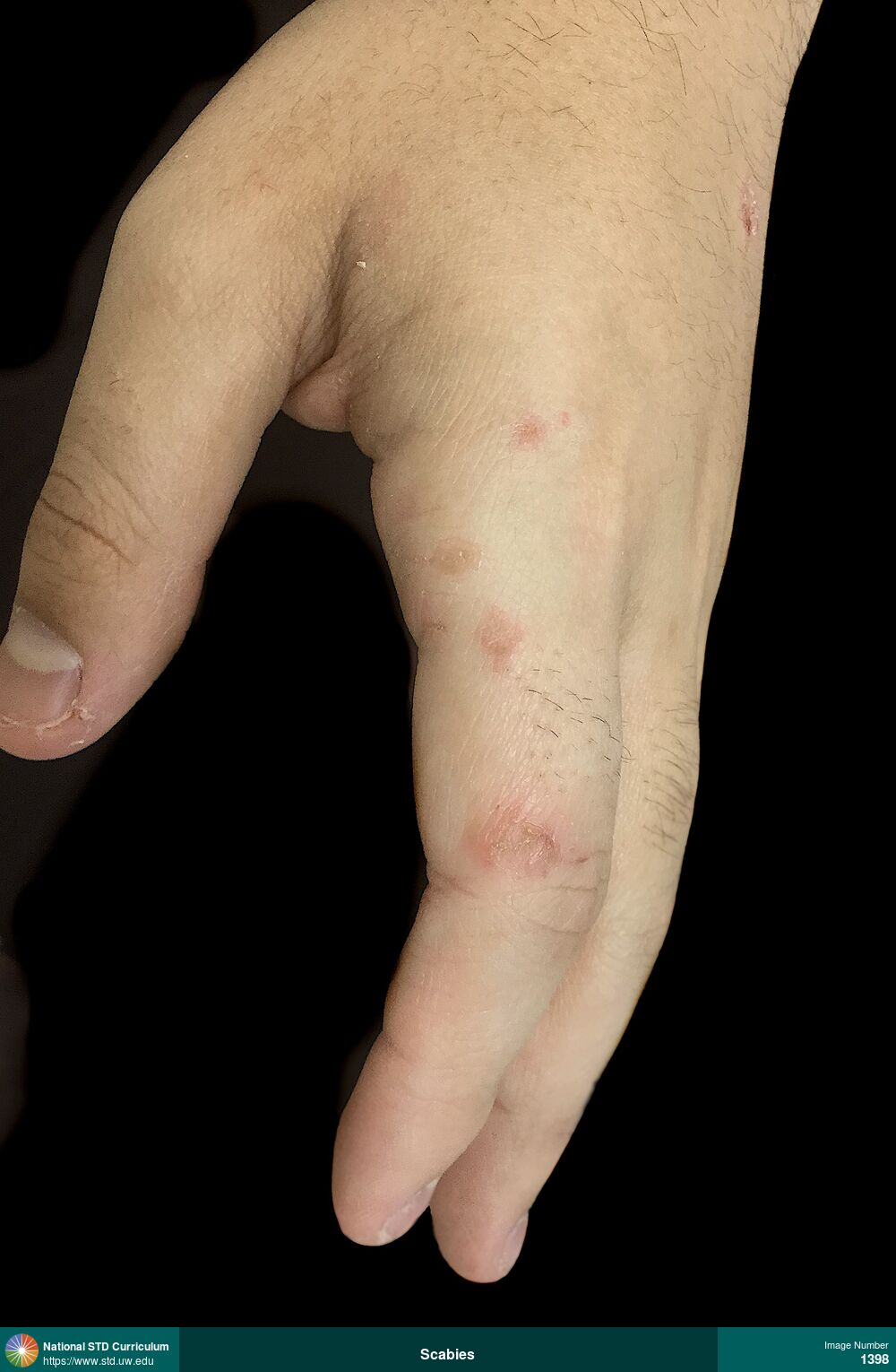
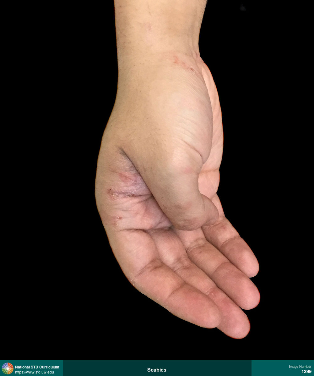
Scabies
Papular, pruritic lesions on the left hand caused by scabies.
Photo: Erythema, Papule / Papules, Hand (Left), Itch
Courtesy of Negusse Ocbamichael, PA
Erythema, Papule / Papules Hand (Left)
1398

Syphilis-Secondary
Hyperpigmented, maculopapular rash on the soles of feet in a man with secondary syphilis.
Photo: Annular, Hyperpigmentation, Macule / Macules, Papule / Papules, Rash, Dark skin tone, Feet/Soles, Non-Painful, Rash
Courtesy of Negusse Ocbamichael, PA
Annular, Hyperpigmentation, Macule / Macules, Papule / Papules, Rash Dark skin tone, Feet/Soles
1404
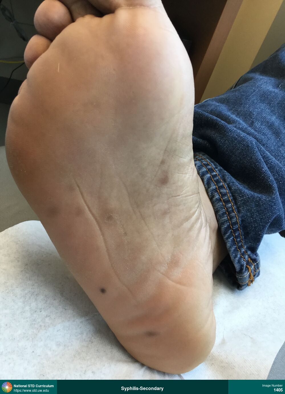
Syphilis-Secondary
Hyperpigmented, maculopapular rash on the soles of feet (right foot shown) in a man with secondary syphilis.
Photo: Annular, Hyperpigmentation, Macule / Macules, Papule / Papules, Rash, Dark skin tone, Feet/Soles, Non-Painful, Rash
Courtesy of Negusse Ocbamichael, PA
Annular, Hyperpigmentation, Macule / Macules, Papule / Papules, Rash Dark skin tone, Feet/Soles
1405
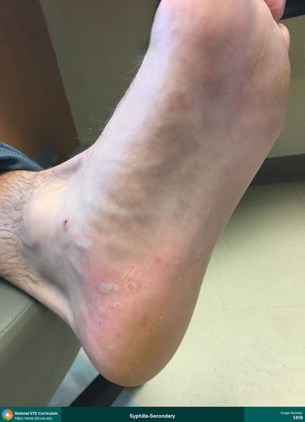
Syphilis-Secondary
Papular and pustular rash on right foot in a man with secondary syphilis.
Photo: Annular, Papule / Papules, Pustule / Pustules, Feet/Soles, Light skin tone, Non-Painful
Courtesy of Negusse Ocbamichael, PA
Annular, Papule / Papules, Pustule / Pustules Feet/Soles, Light skin tone
1419
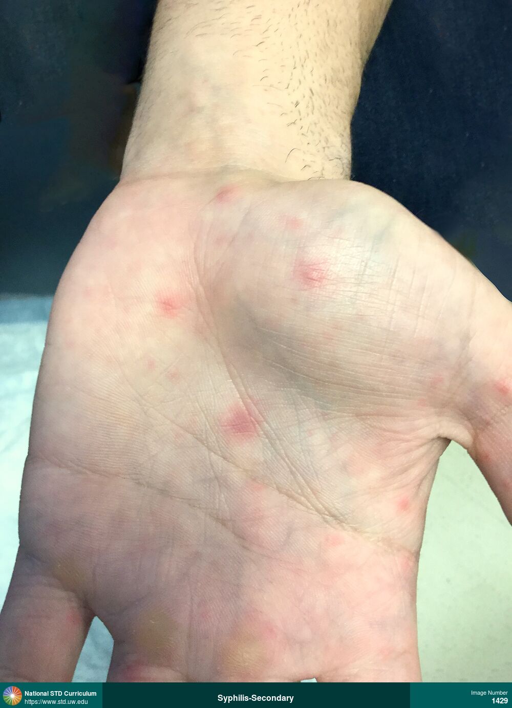
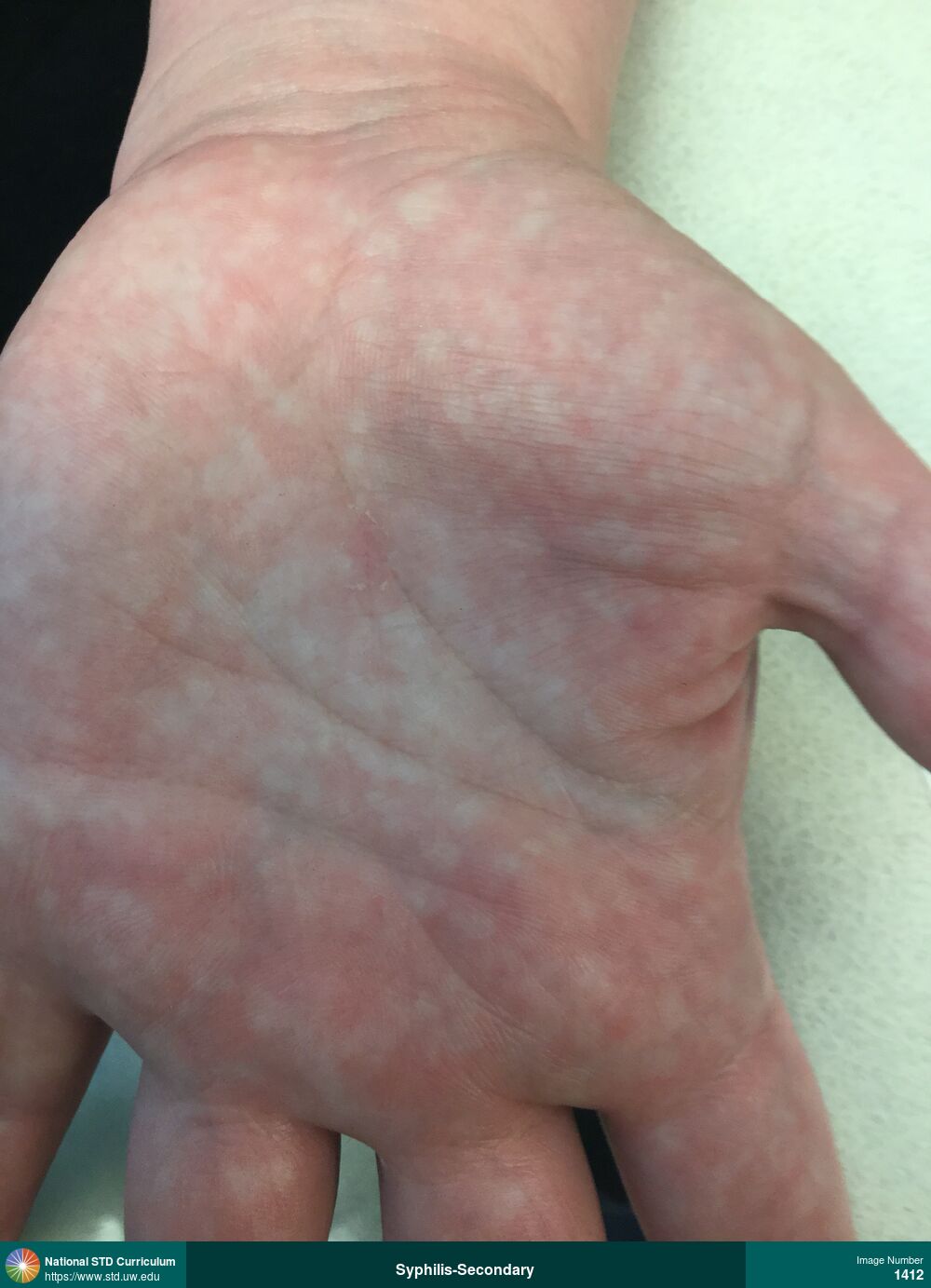
Syphilis-Secondary
Erythematous, maculopapular rash on left palm in a man with secondary syphilis.
Photo: Annular, Erythema, Macule / Macules, Papule / Papules, Arm (Left), Light skin tone, Non-Painful
Courtesy of Negusse Ocbamichael, PA
Annular, Erythema, Macule / Macules, Papule / Papules Arm (Left), Light skin tone
1429
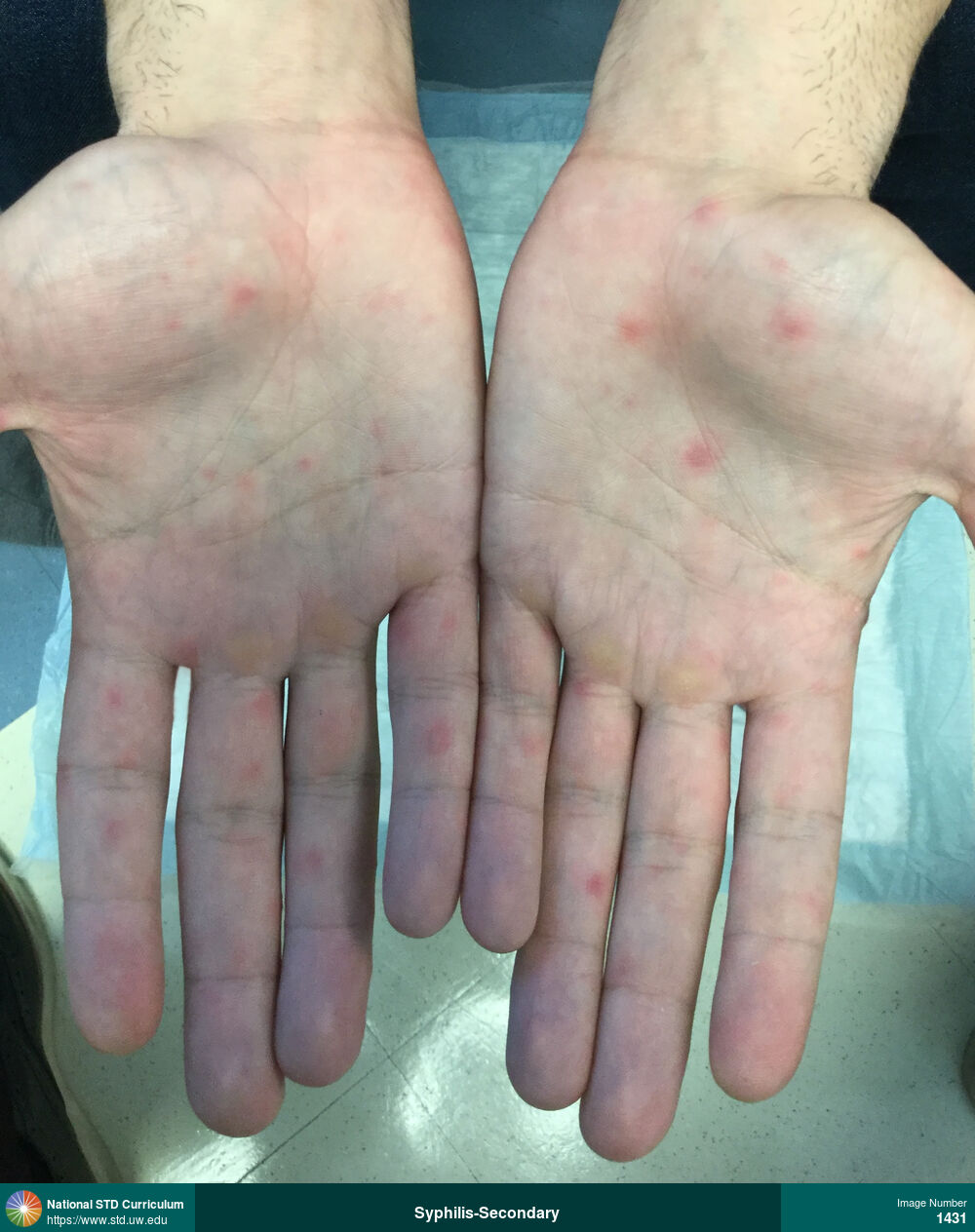
Syphilis-Secondary
Erythematous, maculopapular rash on bilateral palms in a man with secondary syphilis.
Photo: Annular, Erythema, Macule / Macules, Papule / Papules, Hand (Left), Hand (Right), Light skin tone
Courtesy of Negusse Ocbamichael, PA
Annular, Erythema, Macule / Macules, Papule / Papules Hand (Left), Hand (Right), Light skin tone
1431
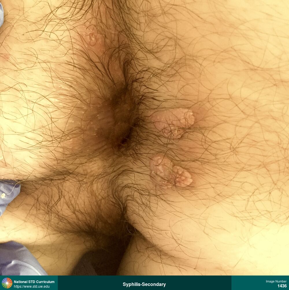
Syphilis-Secondary
Man with two large verrucous plaques on the right perianal region that was confirmed as secondary syphilis and condylomata lata. A sample from one of the lesions, when viewed under Darkfield microscopy, was teeming with spirochetes).
Photo: Papule / Papules, Verrucous, Anal / Perianal, Buttock, Non-Painful
Courtesy of Negusse Ocbamichael, PA
Papule / Papules, Verrucous Anal / Perianal, Buttock
1436
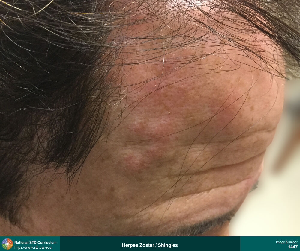
Herpes Zoster / Shingles
Painful (burning sensation), erythematous, vesicular lesions that developed unilaterally in cranial nerve V1 distribution on the right forehead caused by herpes zoster (shingles).
Photo: Erythema, Papule / Papules, Vesicle / Vesicles, Face, Light skin tone, Painful, Rash
Courtesy of Negusse Ocbamichael, PA
Erythema, Papule / Papules, Vesicle / Vesicles Face, Light skin tone
1447
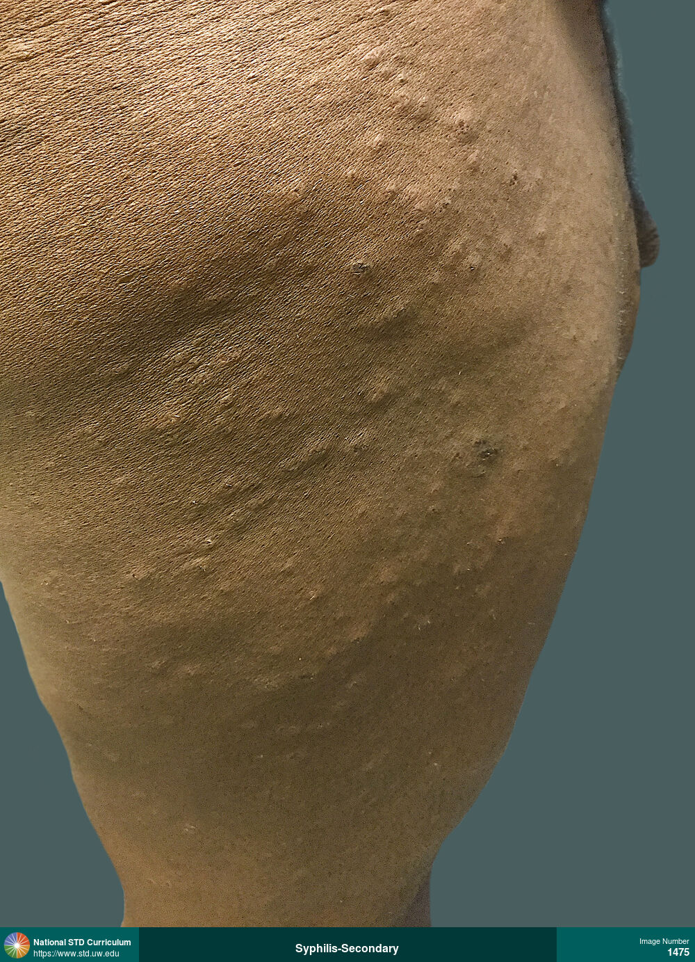
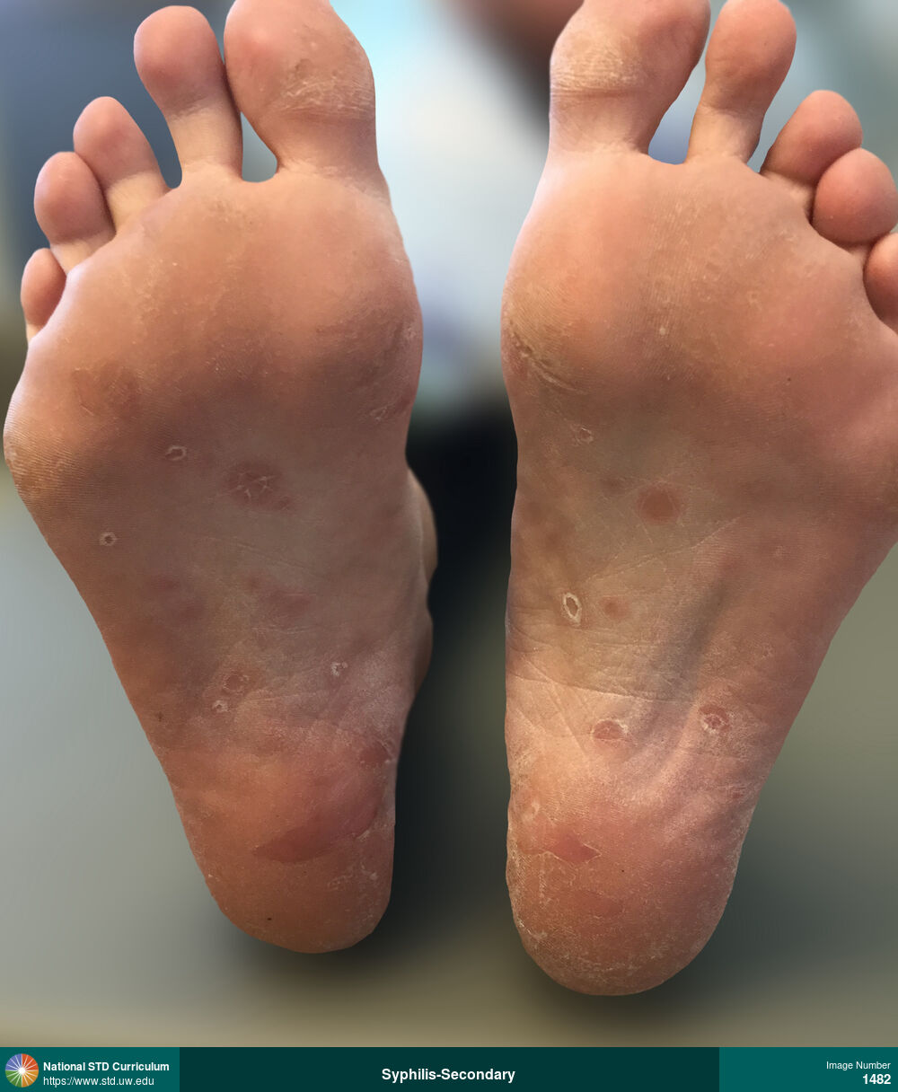
Syphilis-Secondary
Erythematous maculopapular rash on soles in a man with secondary syphilis.
Photo: Annular, Macule / Macules, Papule / Papules, Feet, Non-Painful, Rash
Courtesy of Negusse Ocbamichael, PA
Annular, Macule / Macules, Papule / Papules Feet
1482
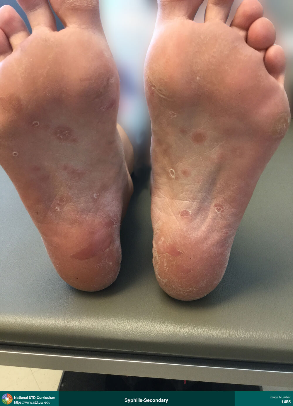
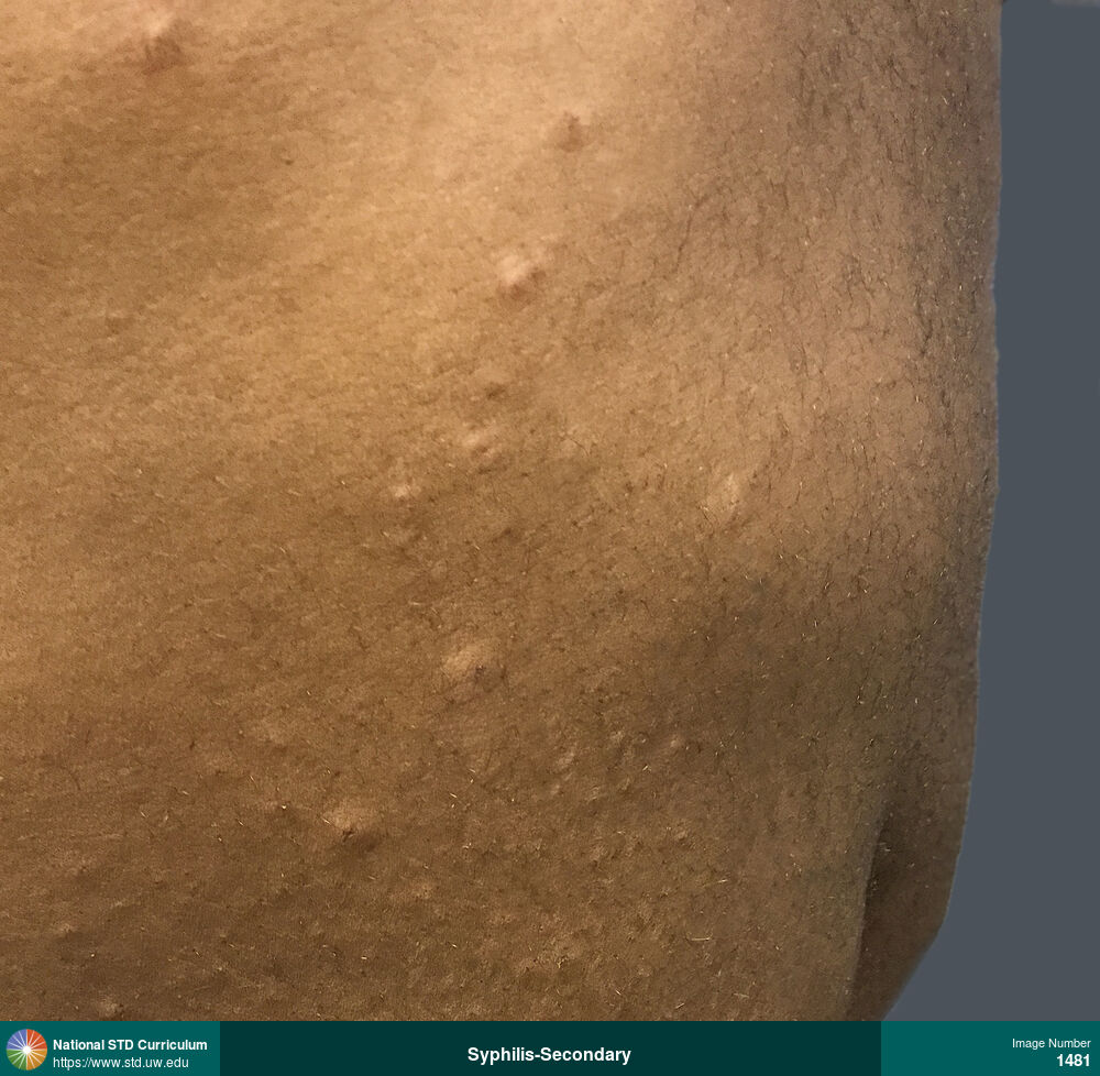
Syphilis-Secondary
Erythematous maculopapular rash on soles in a man with secondary syphilis.
Photo: Annular, Hyperpigmentation, Macule / Macules, Papule / Papules, Feet/Soles, Non-Painful, Rash
Courtesy of Negusse Ocbamichael, PA
Annular, Hyperpigmentation, Macule / Macules, Papule / Papules Feet/Soles
1485
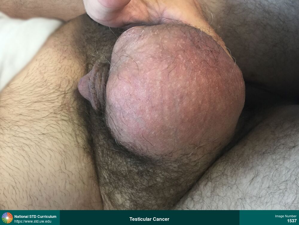
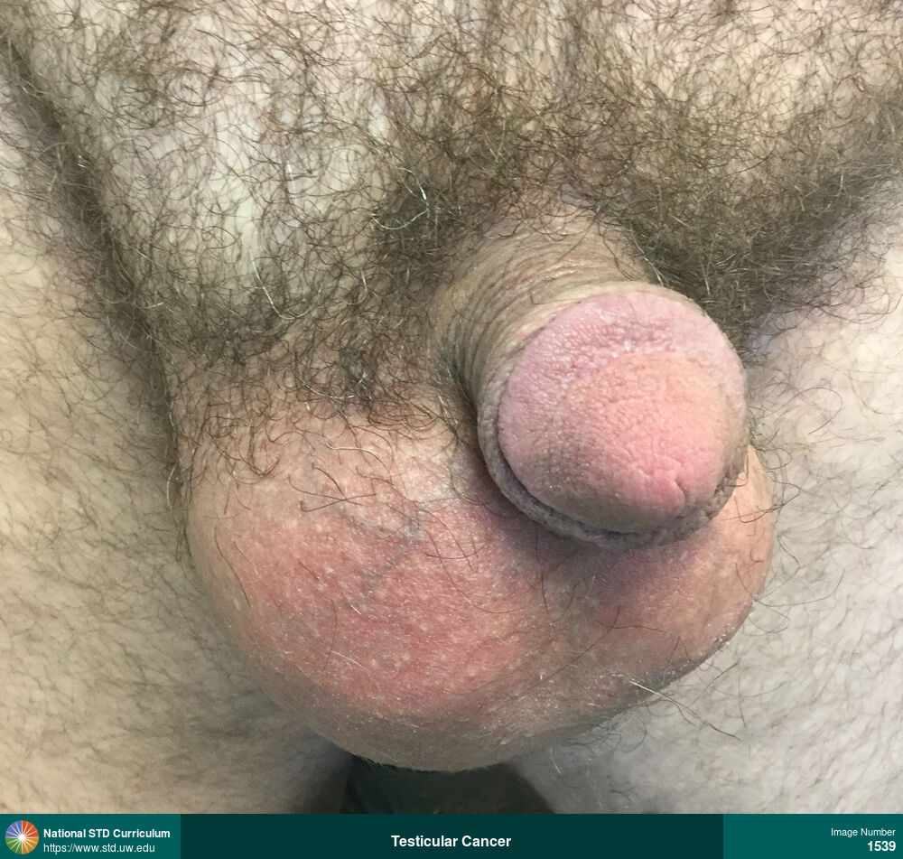
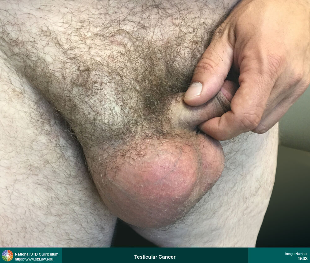
Testicular Cancer
Frontal view of scrotum, testes, and right inguinal region. Examination showed a arge, nontender firm, right testicular mass in a man diagnosed with right-sided testicular cancer. There is also prominent right inguinal adenopathy.
Photo: Adenopathy, Edema / Swelling, Mass, Groin/Inguinal, Light skin tone, Scrotum
Courtesy of Negusse Ocbamichael, PA
Adenopathy, Edema / Swelling, Mass Groin/Inguinal, Light skin tone, Scrotum
1543

Tinea cruris
Erythematous, annular, macular rash with well-defined borders in suprapubic and inguinal regions that was diagnosed as tinea cruris.
Photo: Annular, Erythema, Patch/Patches, Rash, Groin/Inguinal, Light skin tone, Suprapubic (Hypogastrium), Itch, Non-Painful
Courtesy of Negusse Ocbamichael, PA
Annular, Erythema, Patch/Patches, Rash Groin/Inguinal, Light skin tone, Suprapubic (Hypogastrium)
1553
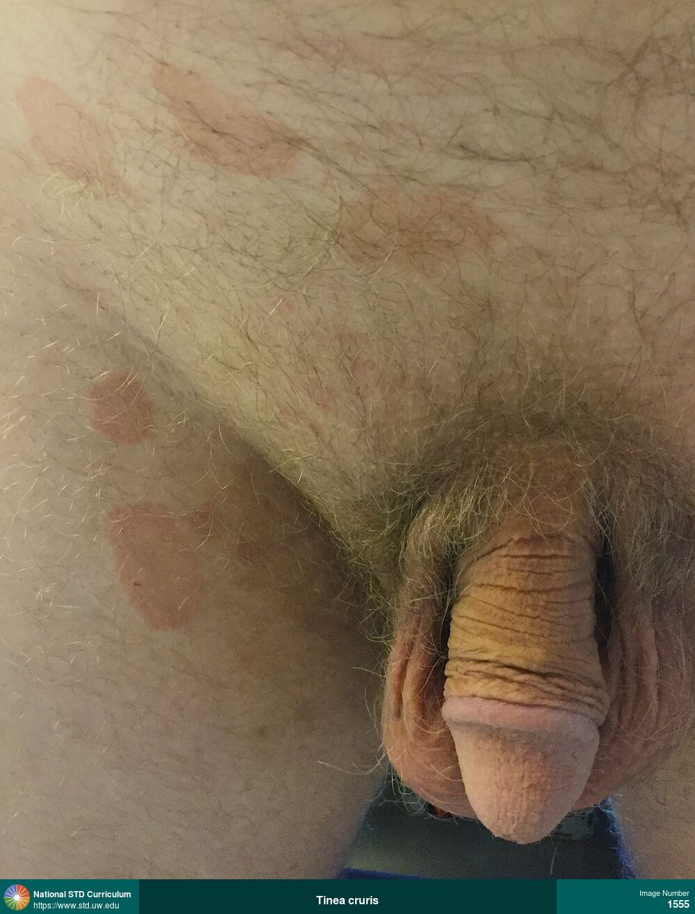
Tinea cruris
Erythematous, annular, macular rash with well-defined borders in suprapubic and inguinal regions that was diagnosed as tinea cruris.
Photo: Annular, Erythema, Patch/Patches, Rash, Groin/Inguinal, Light skin tone, Suprapubic (Hypogastrium), Itch, Non-Painful, Rash
Courtesy of Negusse Ocbamichael, PA
Annular, Erythema, Patch/Patches, Rash Groin/Inguinal, Light skin tone, Suprapubic (Hypogastrium)
1555
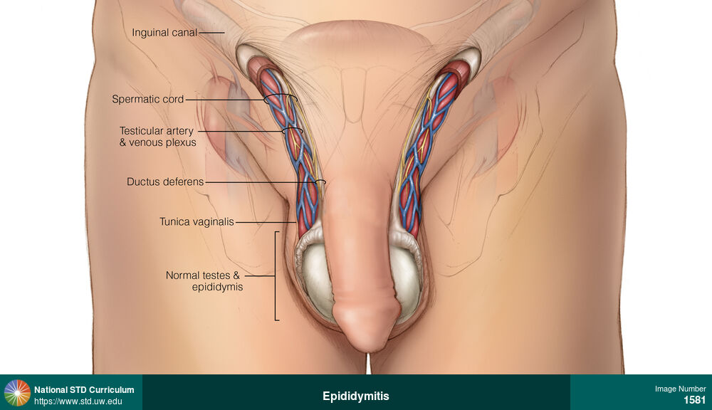
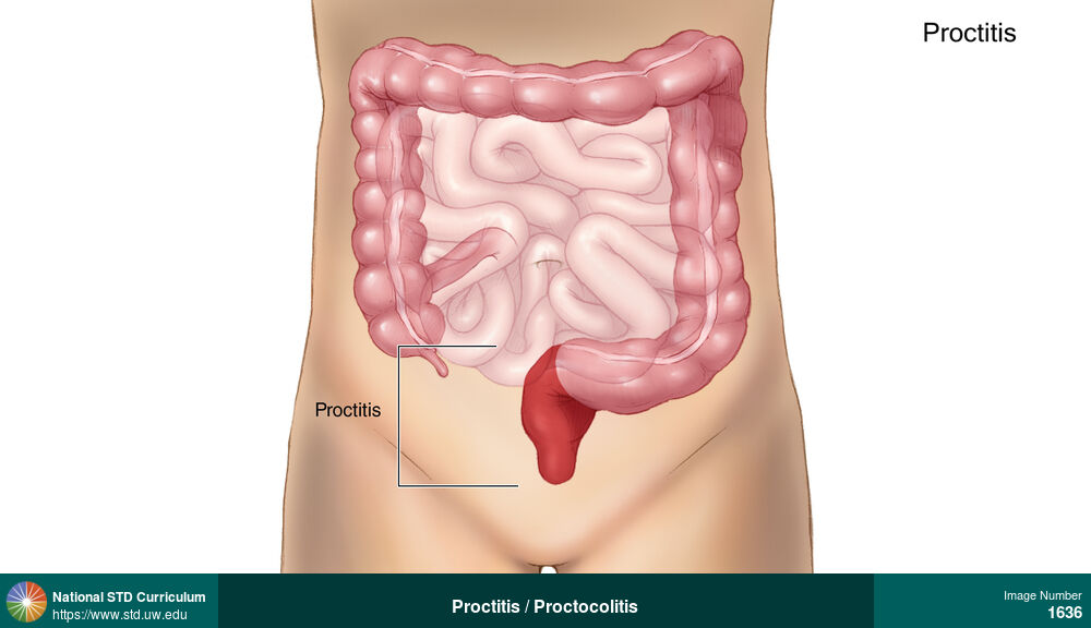
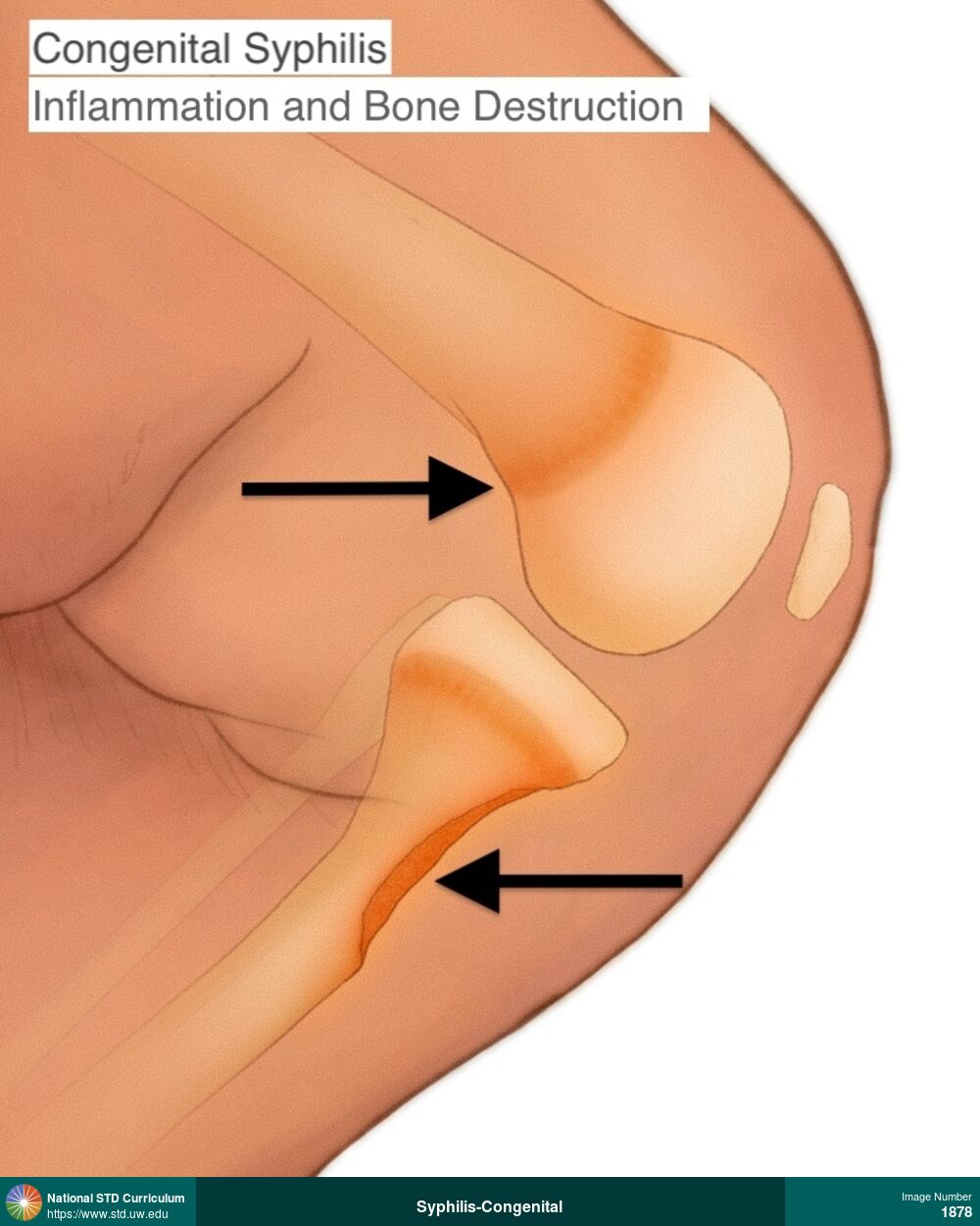
Epididymitis
This illustration shows normal appearance of epididymis, testicle, and scrotal sac. The epididymis is a coiled tube Ton the back of the testicle and it functions to store and transport sperm.
Illustration: Epididymis, Penis, Scrotum
Courtesy of Cognition Studio, Inc. and David H. Spach, MD
N/A Epididymis, Penis, Scrotum
1581
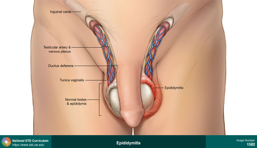
Epididymitis
This illustration shows characteristic inflammation and swelling of the left epididymis and left scrotal sac. The epididymis is a coiled tube located on the back of the testicle, and it functions to store and transport sperm.
Illustration: Edema / Swelling, Epididymis, Penis, Scrotum, Painful
Courtesy of Cognition Studio, Inc. and David H. Spach, MD
Edema / Swelling Epididymis, Penis, Scrotum
1582
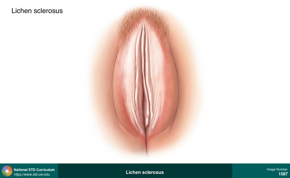
Lichen sclerosus
This illustration of lichen sclerosus on the vulva shows smooth, flat, hypopigmented, thinning patches bilaterally, with extensive involvement on both the labia majora and labia minora.
Illustration: Hypopigmentation, Patch/Patches, Light skin tone, Vulva, Itch
Courtesy of Cognition Studio, Inc. and David H. Spach, MD
Hypopigmentation, Patch/Patches Light skin tone, Vulva
1587
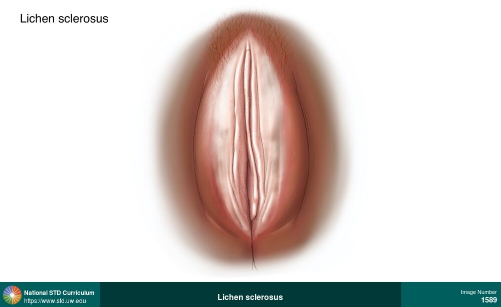
Lichen sclerosus
This illustration of lichen sclerosus on the vulva shows smooth, flat, hypopigmented, thinning patches bilaterally, with extensive involvement on both the labia majora and labia minora.
Illustration: Hypopigmentation, Patch/Patches, Dark skin tone, Vulva, Itch
Courtesy of Cognition Studio, Inc. and David H. Spach, MD
Hypopigmentation, Patch/Patches Dark skin tone, Vulva
1589
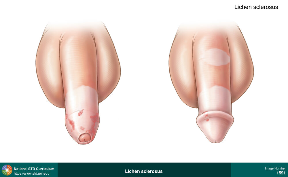
Lichen sclerosus
This illustration of lichen sclerosus on the penis shows several white patches of thinned skin on uncircumcised penis (on the left) and circumcised penis (on the right). The foreskin has been constricted (tightened) by this process.
Illustration: Hypopigmentation, Plaque, Light skin tone, Penis, Itch
Courtesy of Cognition Studio, Inc. and David H. Spach, MD
Hypopigmentation, Plaque Light skin tone, Penis
1591
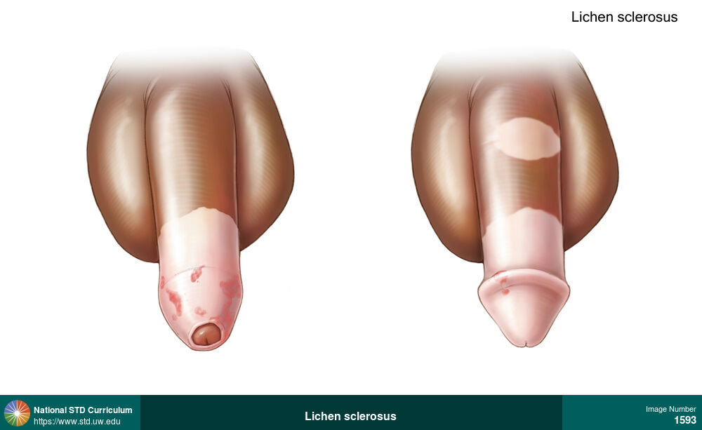
Lichen sclerosus
This illustration of lichen sclerosus on the penis shows several white patches of thinned skin on uncircumcised penis (on the left) and circumcised penis (on the right). The foreskin has been constricted (tightened) by this process.
Illustration: Hypopigmentation, Plaque, Dark skin tone, Penis, Itch
Courtesy of Cognition Studio, Inc. and David H. Spach, MD
Hypopigmentation, Plaque Dark skin tone, Penis
1593
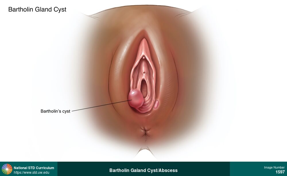
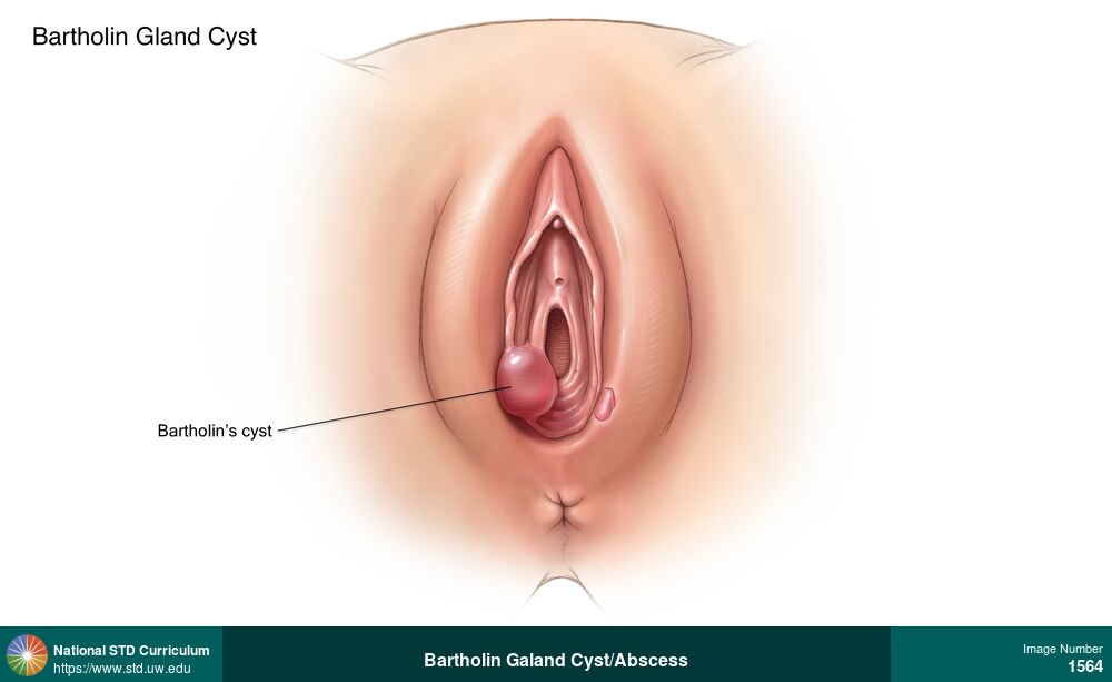
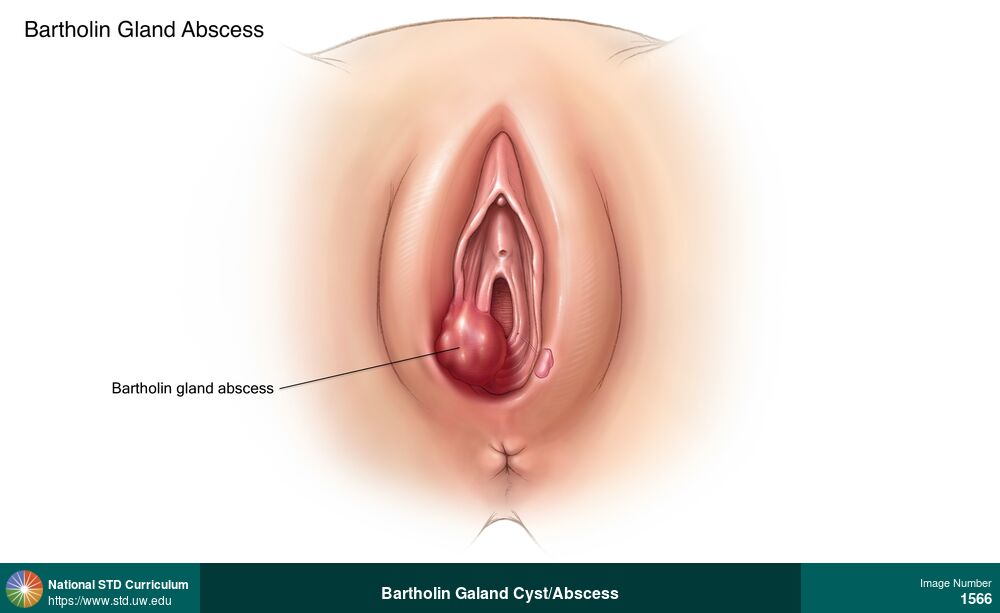
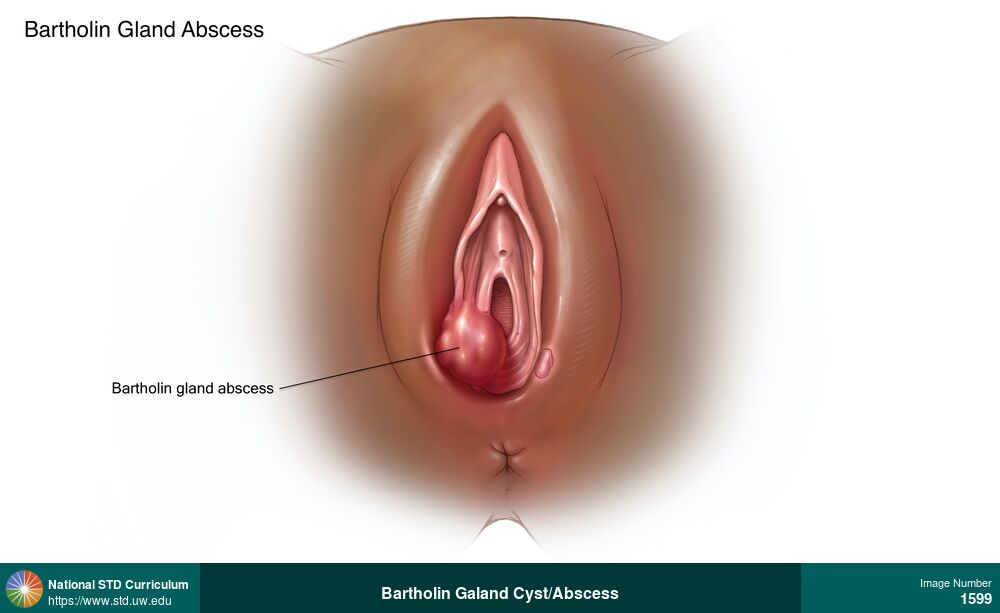
Bartholin Galand Cyst/Abscess
A Bartholin gland cyst is caused by increased fluid collection in the Bartholin’s glands that develops after an obstruction in the Bartholin’s duct. A Bartholin gland cyst manifests clinically as a firm, swollen, fluid-filled lump in the tissues at the lower opening of the vagina. The cyst and tissues surrounding the cyst may become erythematous and/or tender.
Illustration: Cyst, Edema / Swelling, Erythema, Dark skin tone, Vulva
Courtesy of Cognition Studio, Inc. and David H. Spach, MD
Cyst, Edema / Swelling, Erythema Dark skin tone, Vulva
1597
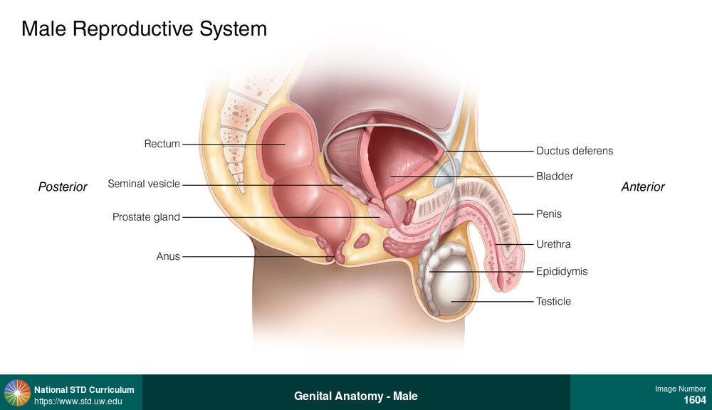

Genital Anatomy - Male
Frontal view of male reproductive system, including circumcised penis, epididymis, testes, spermatic cord, and tunica vaginalis.
Illustration: Epididymis, Light skin tone, Penis, Scrotum
Courtesy of Cognition Studio, Inc. and David H. Spach, MD
N/A Epididymis, Light skin tone, Penis, Scrotum
1605

Testicular Torsion
This illustration of a young man with testicular torsion shows apparent twisting of the spermatic cord and reduced blood flow to the left testicle. The left testicle appears slighty elevated in the scrotum compared to the right testicle.
Illustration: Edema / Swelling, Epididymis, Light skin tone, Scrotum, Painful
Courtesy of Cognition Studio, Inc. and David H. Spach, MD
Edema / Swelling Epididymis, Light skin tone, Scrotum
1606
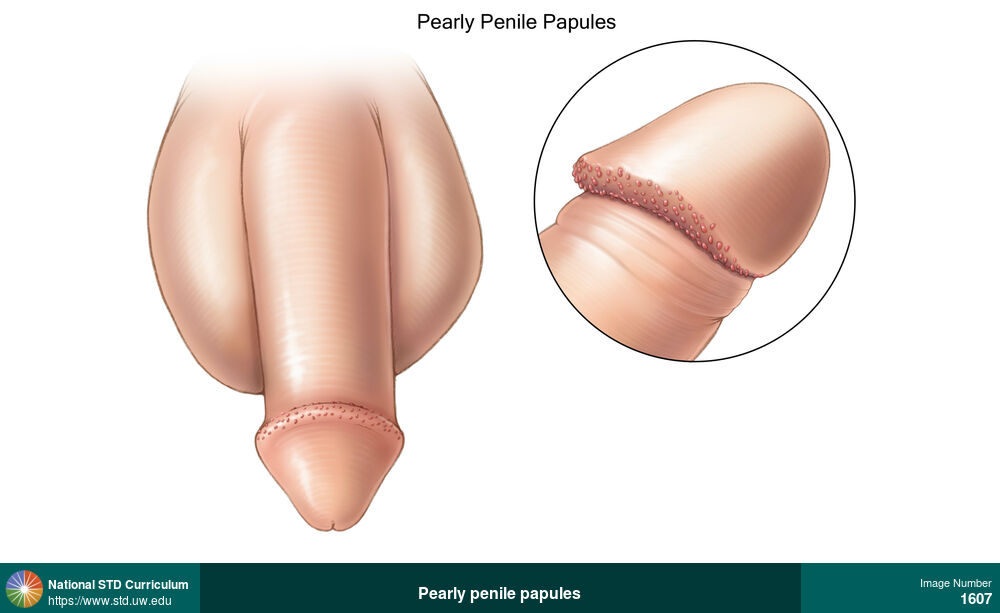
Pearly penile papules
Pearly penile papules are a normal anatomic variant that appear as small (<1 mm), non-tender, flesh-colored bumps in rows around the corona of the penis.
Illustration: Papule / Papules, Light skin tone, Penis, Non-Itchy, Non-Painful
Courtesy of Cognition Studio, Inc. and David H. Spach, MD
Papule / Papules Light skin tone, Penis
1607
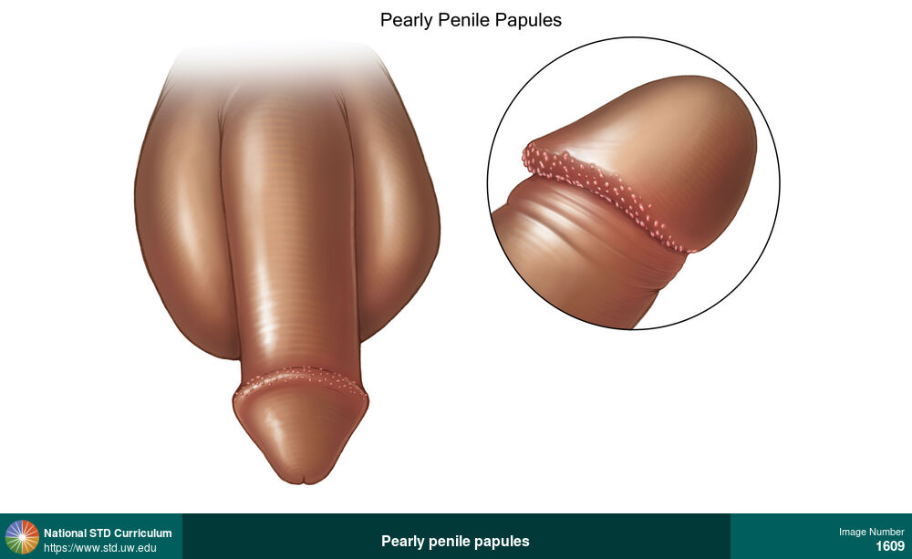
Pearly penile papules
Pearly penile papules are a normal anatomic variant that appear as small (<1 mm), non-tender, flesh-colored bumps in rows around the corona of the penis.
Illustration: Papule / Papules, Dark skin tone, Penis, Non-Itchy, Non-Painful
Courtesy of Cognition Studio, Inc. and David H. Spach, MD
Papule / Papules Dark skin tone, Penis
1609
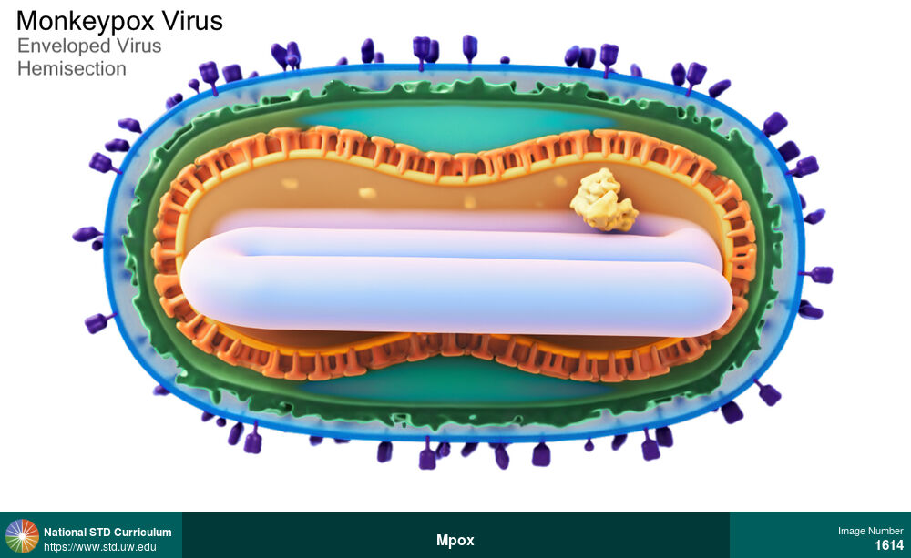
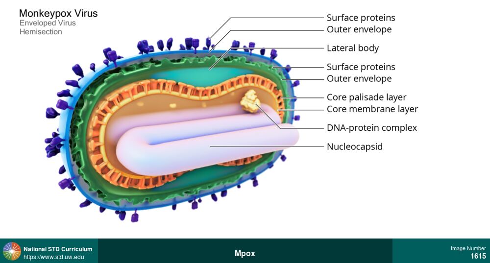

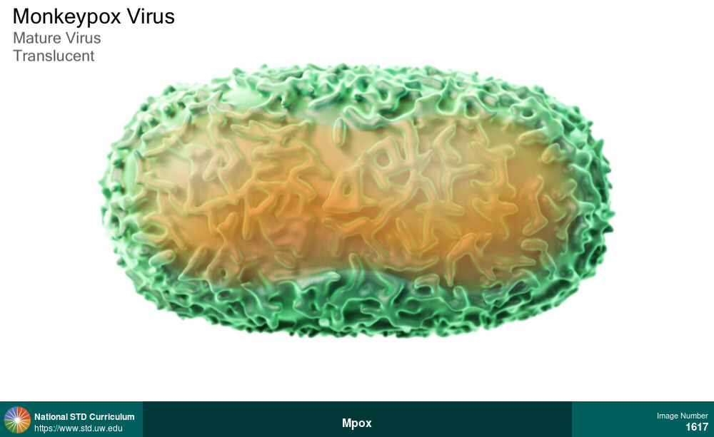
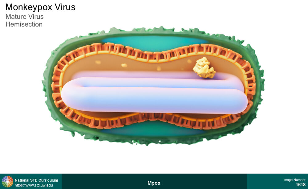
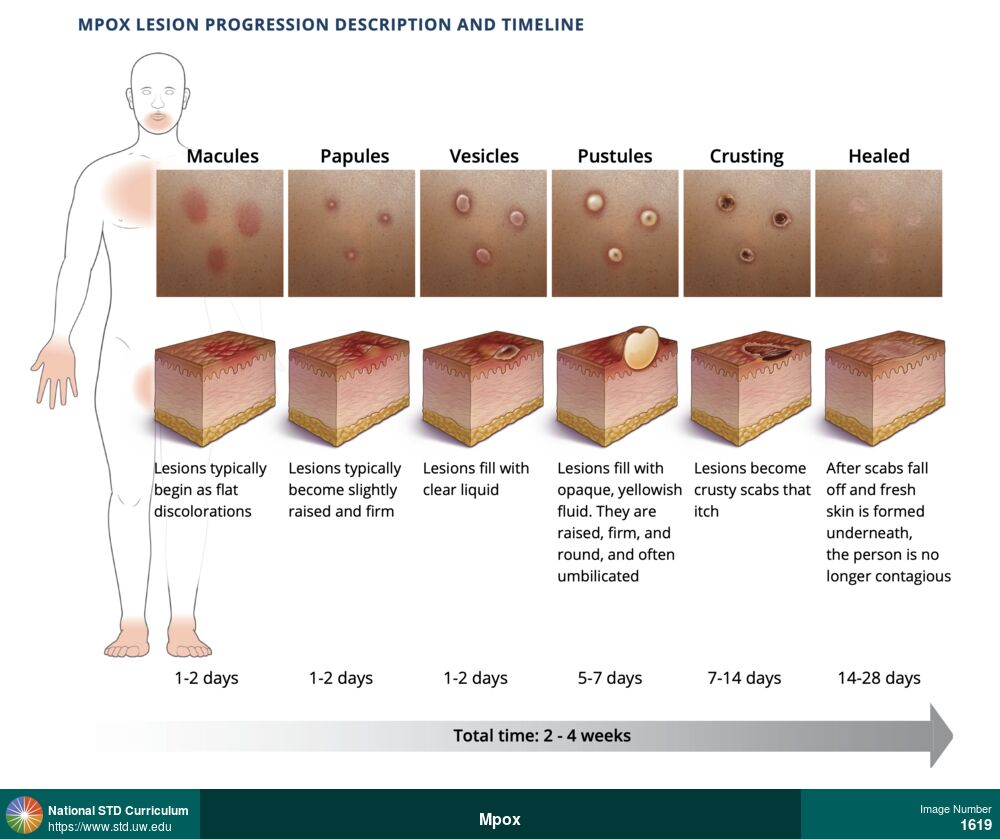
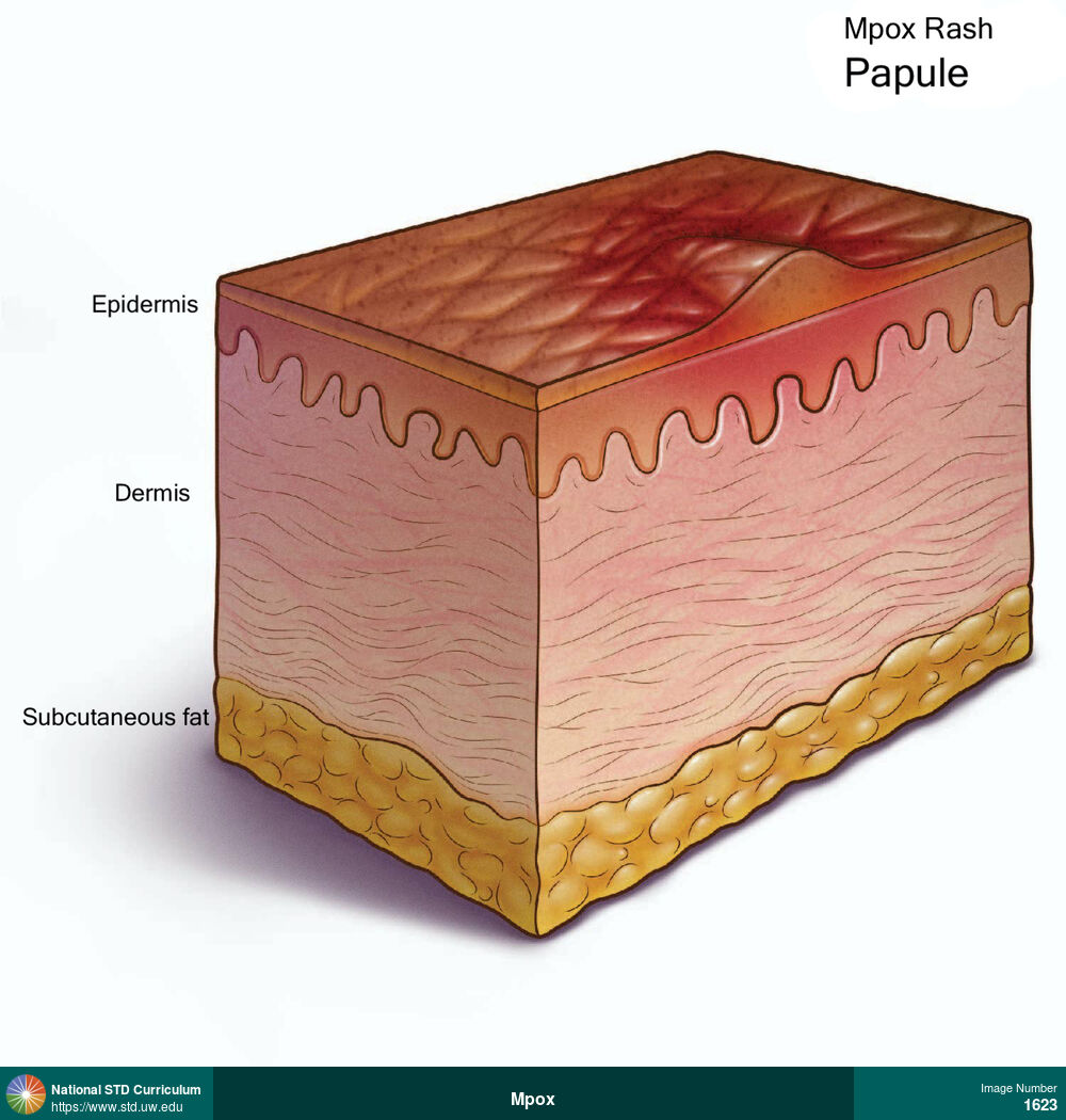
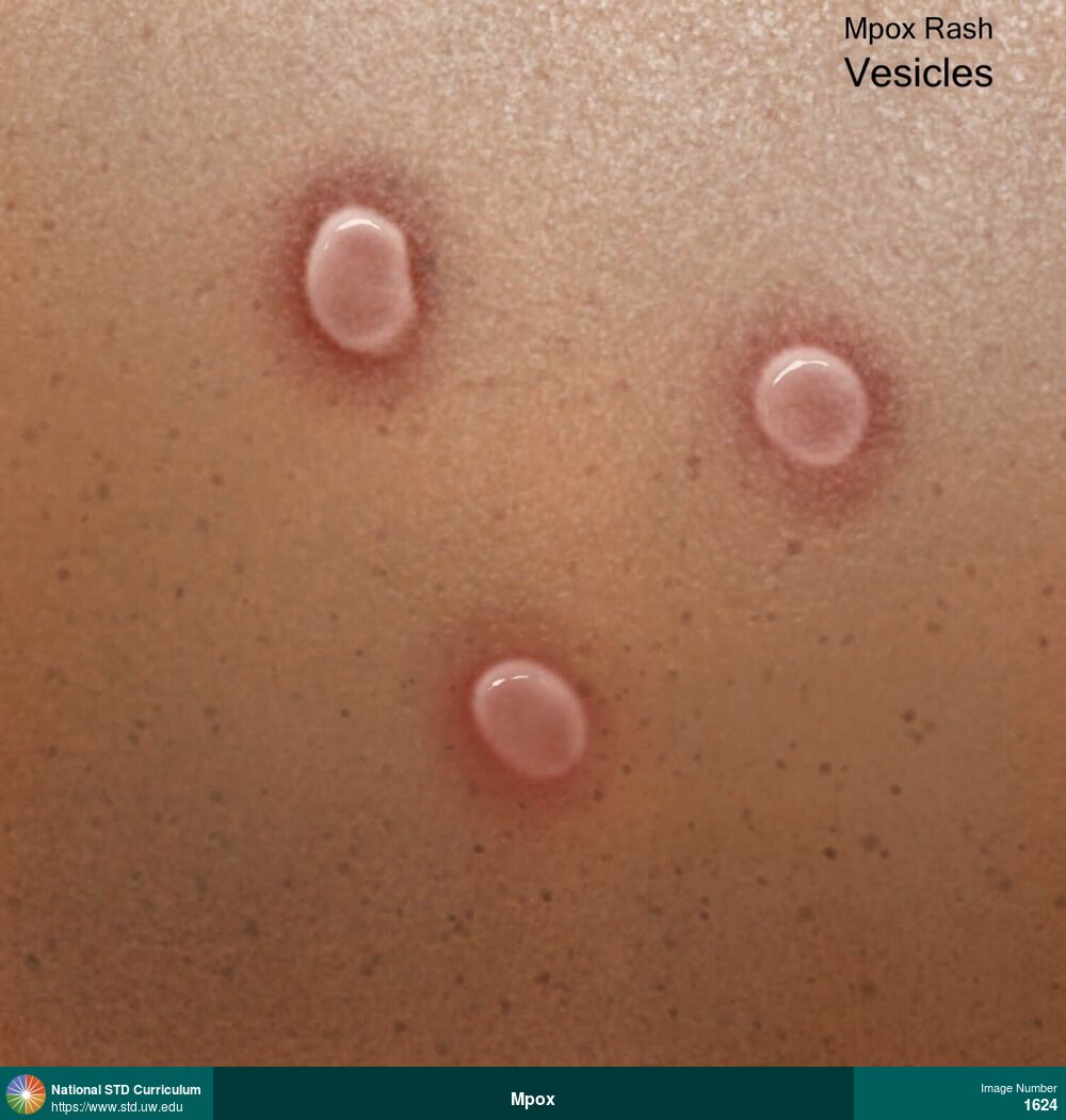
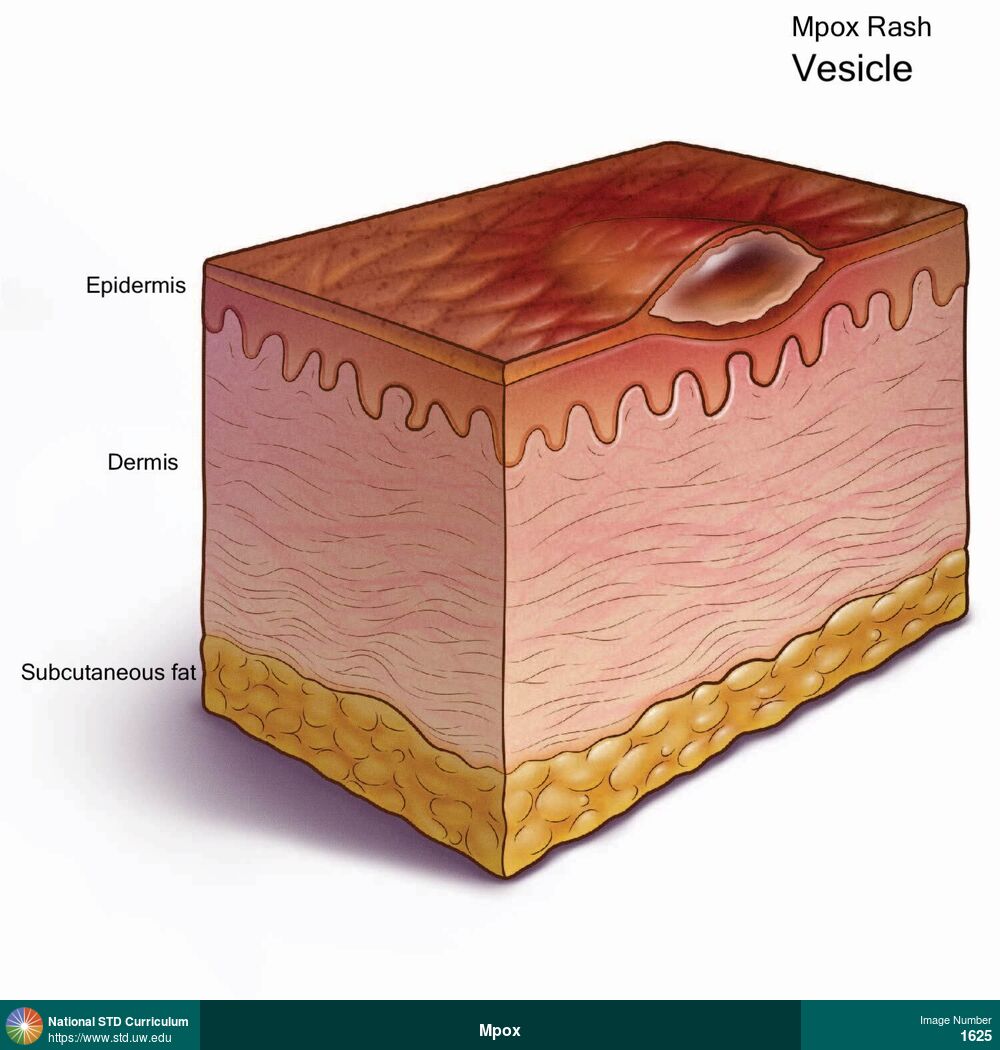
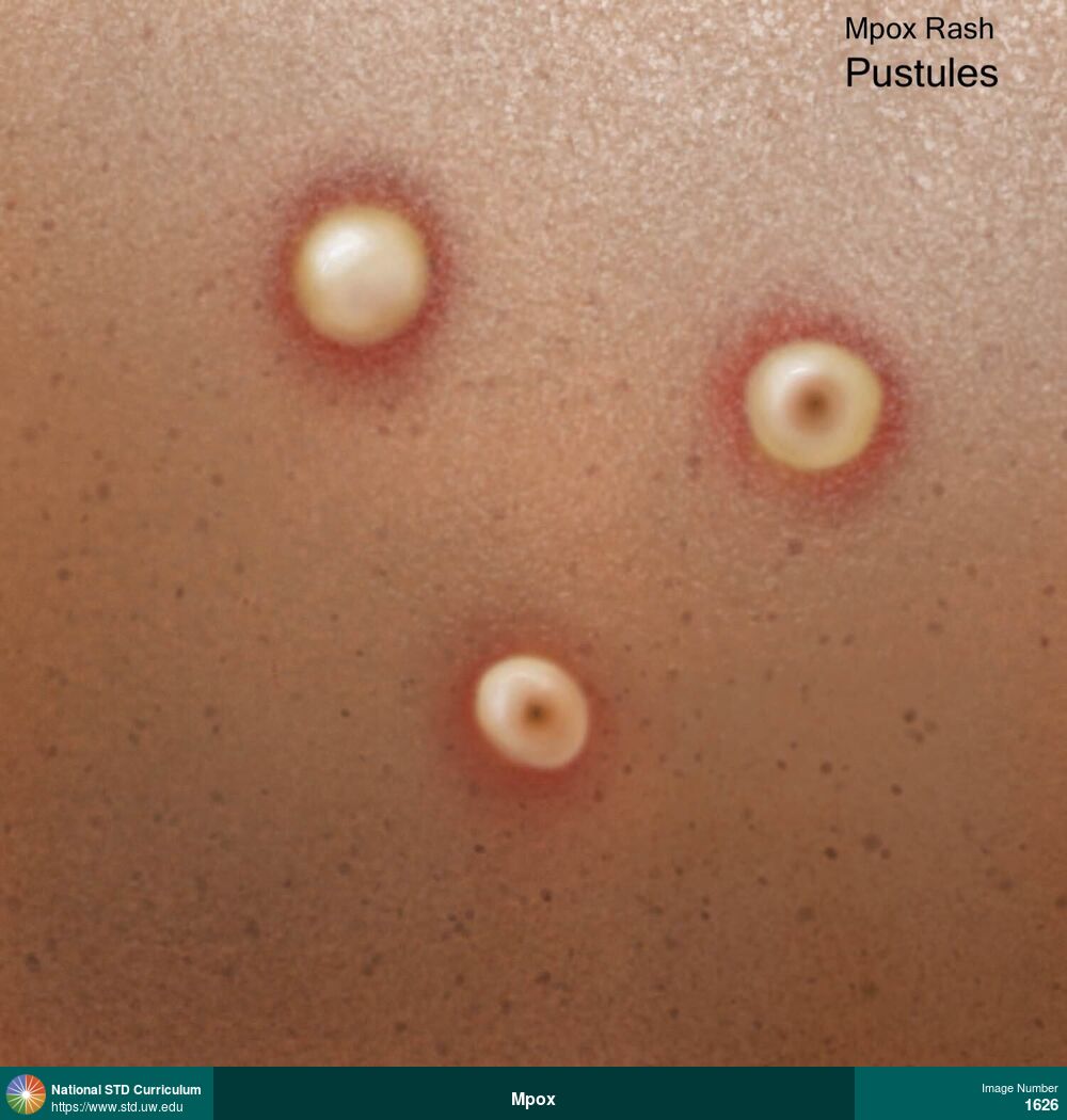
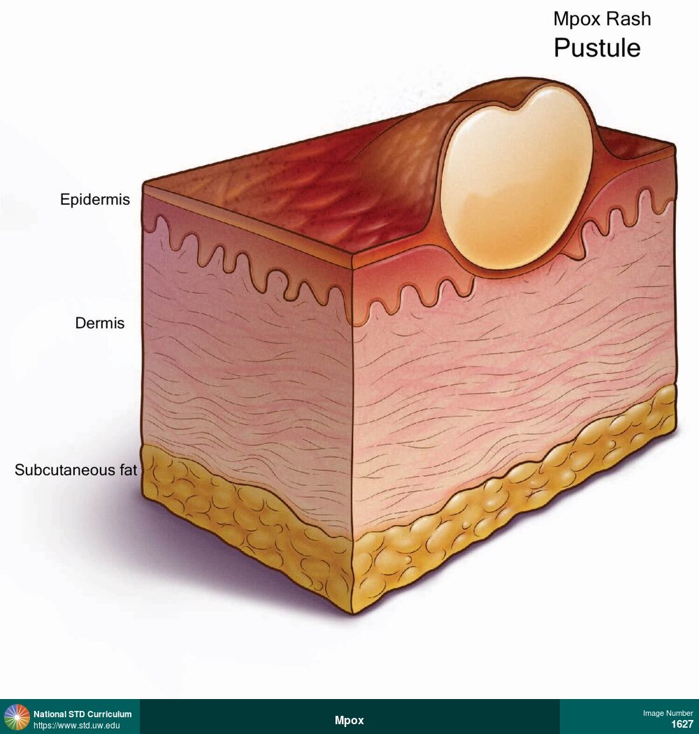
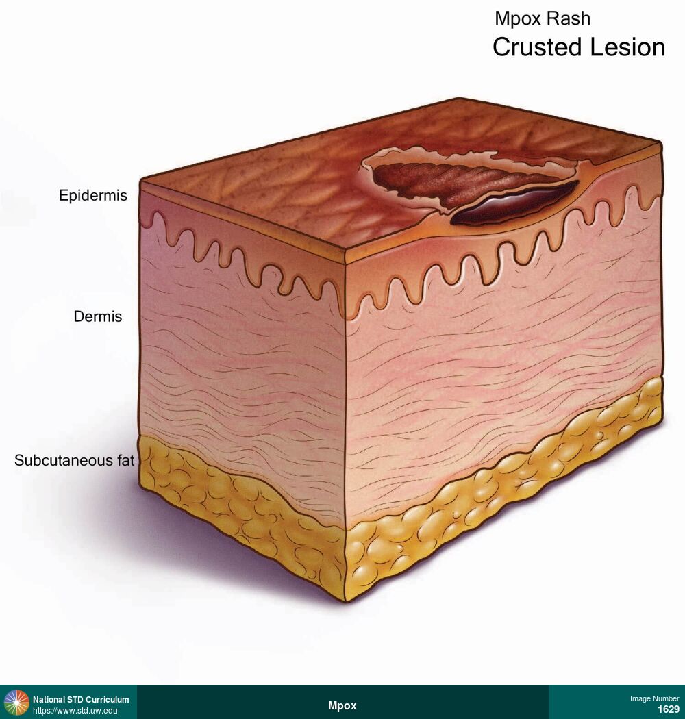
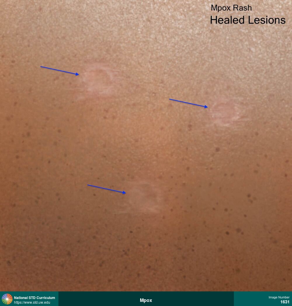
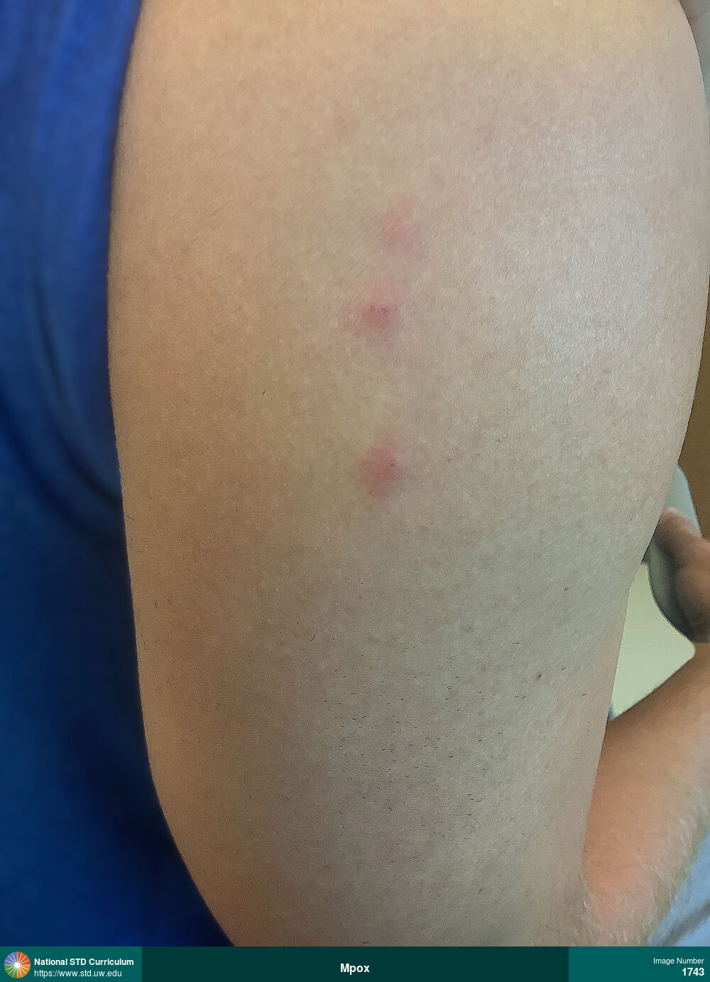
Mpox
Mpox lesions typically progress through six stages shown in this illustration: macules, papules, vesicles, pustules, crusting, and healing.
Illustration: Macule / Macules, Papule / Papules, Pustule / Pustules, Rash, Scab, Ulcer / Ulcers, Vesicle / Vesicles
Courtesy of Cognition Studio, Inc. and David H. Spach, MD
Macule / Macules, Papule / Papules, Pustule / Pustules, Rash, Scab, Ulcer / Ulcers, Vesicle / Vesicles N/A
1619


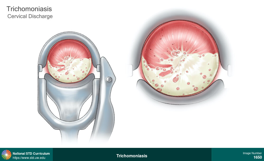
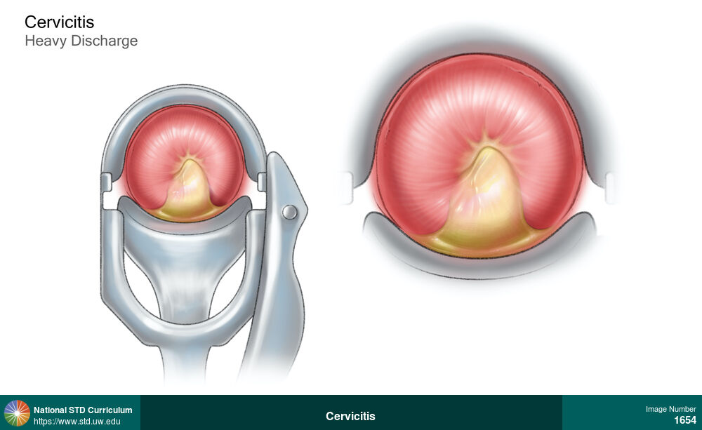
Cervicitis
Speculum view of cervix (left) and close-up view of cervix (right) in a woman with cervicitis showing heavy purulent discharge from the cervical os.
Illustration: Discharge / Vagina / Cervix, Cervix, Discharge
Courtesy of Cognition Studio, Inc. and David H. Spach, MD
Discharge / Vagina / Cervix Cervix
1654
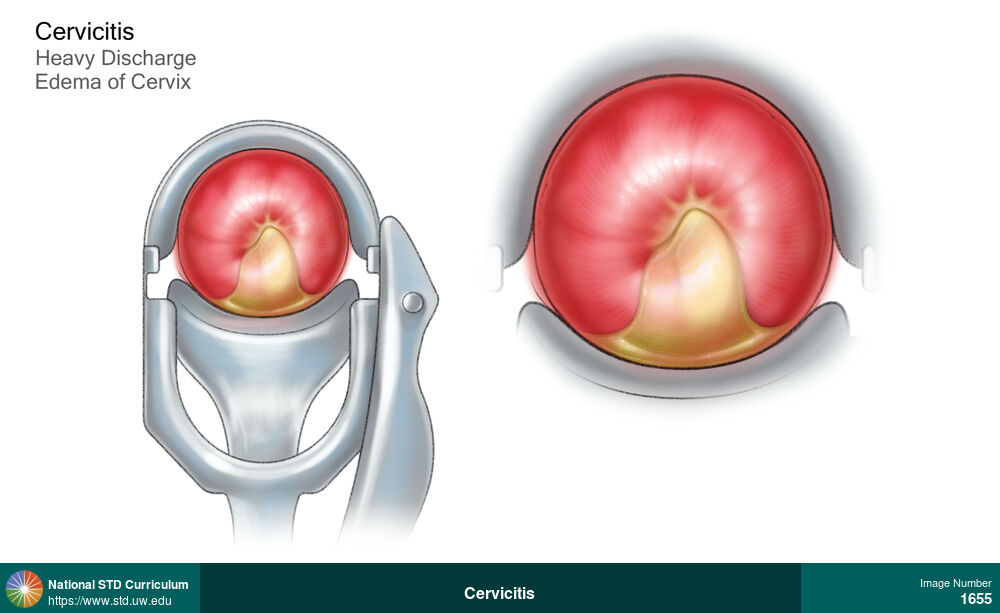
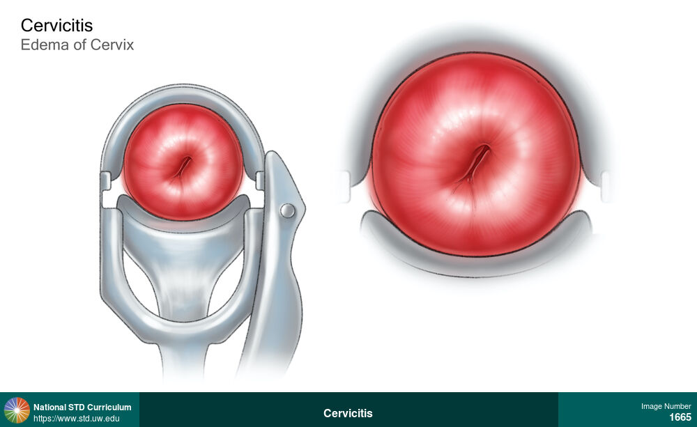
Cervicitis
Speculum view of cervix (left) and close-up view of cervix (right) in a woman with cervicitis showing heavy purulent discharge from the cervical os and edema of cervix.
Illustration: Discharge / Vagina / Cervix, Edema / Swelling, Cervix, Discharge
Courtesy of Cognition Studio, Inc. and David H. Spach, MD
Discharge / Vagina / Cervix, Edema / Swelling Cervix
1655
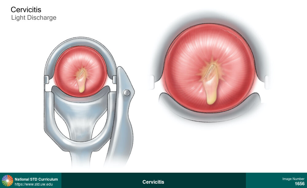
Cervicitis
Speculum view of cervix (left) and close-up view of cervix (right) in a woman with cervicitis showing light (thin) purulent discharge from the cervical os.
Illustration: Discharge / Vagina / Cervix, Cervix, Discharge
Courtesy of Cognition Studio, Inc. and David H. Spach, MD
Discharge / Vagina / Cervix Cervix
1656
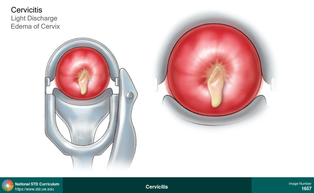
Cervicitis
Speculum view of cervix (left) and close-up view of cervix (right) in a woman with cervicitis showing thin (light) purulent discharge from the cervical os and edema of cervix.
Illustration: Discharge / Vagina / Cervix, Edema / Swelling, Cervix, Discharge
Courtesy of Cognition Studio, Inc. and David H. Spach, MD
Discharge / Vagina / Cervix, Edema / Swelling Cervix
1657
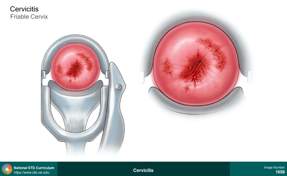
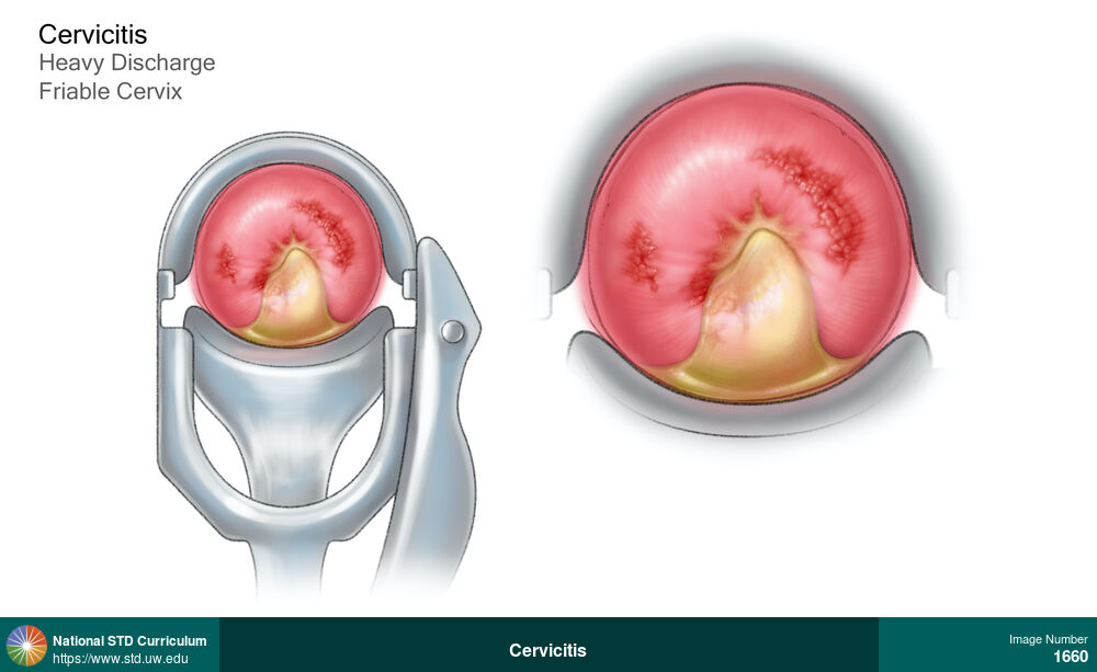
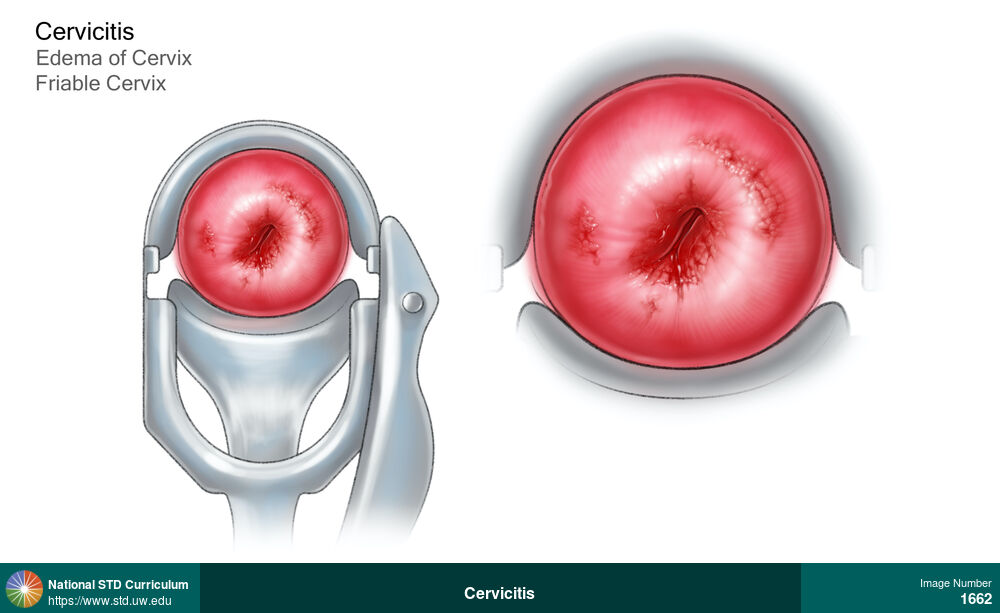
Cervicitis
Speculum view of cervix (left) and close-up view of cervix (right) in a woman with cervicitis showing heavy purulent discharge from the cervical os and friable cervix.
Illustration: Discharge / Vagina / Cervix, Cervix, Discharge
Courtesy of Cognition Studio, Inc. and David H. Spach, MD
Discharge / Vagina / Cervix Cervix
1660
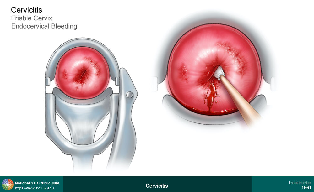
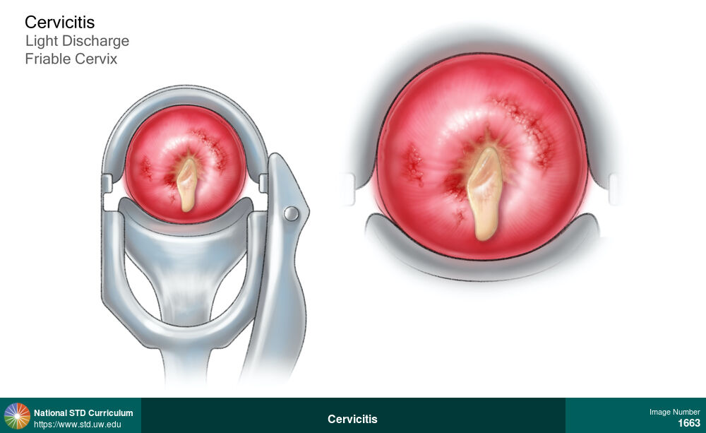
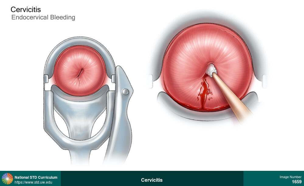
Cervicitis
Speculum view of cervix (left) and close-up view of cervix (right) in a woman with cervicitis showing light (thin) discharge from the cervical os and a friable cervix.
Illustration: Discharge / Vagina / Cervix, Cervix
Courtesy of Cognition Studio, Inc. and David H. Spach, MD
Discharge / Vagina / Cervix Cervix
1663
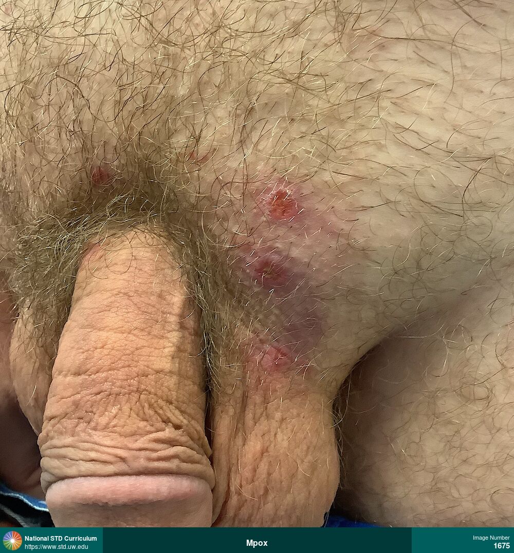
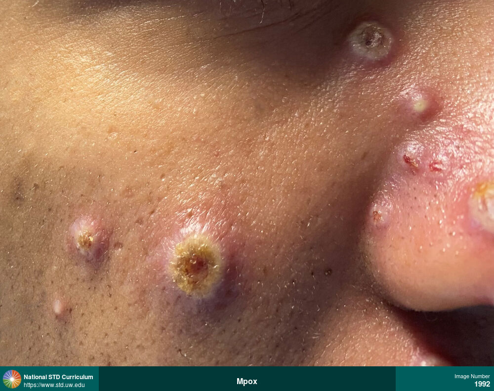
Mpox
Painful, prurtitic, erythematous, with superficial layer of ulceration caused by mpox. These lesions are beginning to heal.
Photo: Erythema, Ulcer / Ulcers, Scrotum, Suprapubic (Hypogastrium), Itch, Painful
Courtesy of Negusse Ocbamichael, PA
Erythema, Ulcer / Ulcers Scrotum, Suprapubic (Hypogastrium)
1675

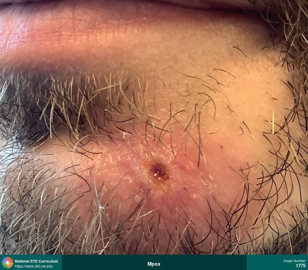
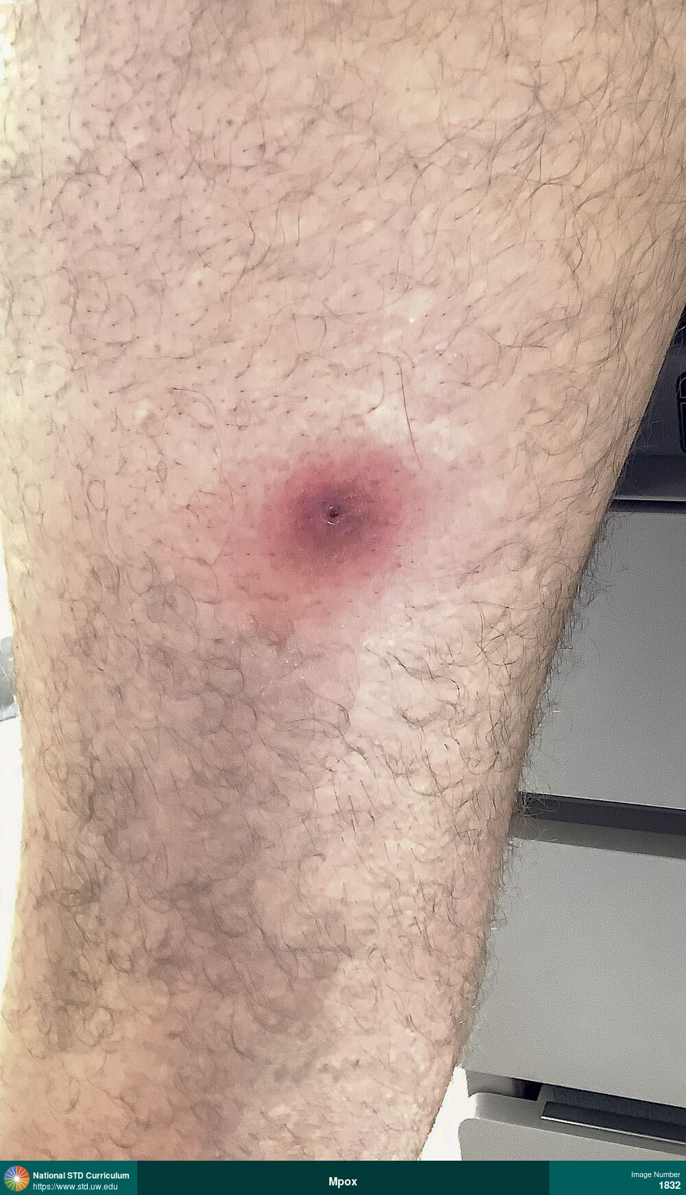
Mpox
Nodular, ulcerative, dome-shaped pustular lesions on the base of the penis and in the suprapubic region just above the scrotum. These lesions, which have a dark core in the center, were confirmed as mpox. The left inguinal node is mildly enlarged.
Photo: Erythema, Pustule / Pustules, Ulcer / Ulcers, Light skin tone, Penis, Scrotum, Suprapubic (Hypogastrium), Painful
Courtesy of Negusse Ocbamichael, PA
Erythema, Pustule / Pustules, Ulcer / Ulcers Light skin tone, Penis, Scrotum, Suprapubic (Hypogastrium)
1685
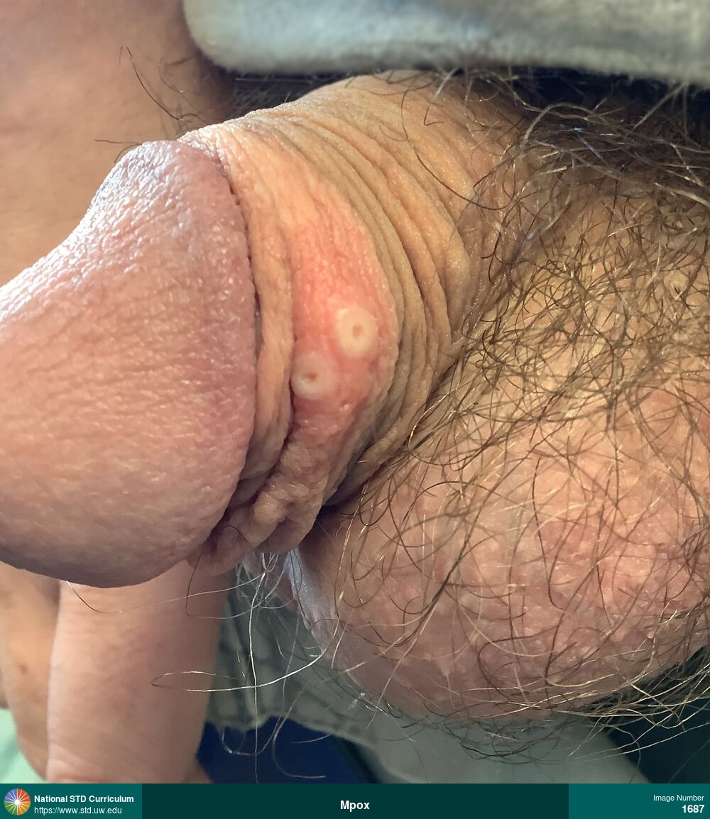
Mpox
Two round papular-pustular lesions with central crater on shaft of penis in a man with mpox.
Photo: Annular, Papule / Papules, Pustule / Pustules, Light skin tone, Penis, Painful, Rash
Courtesy of Negusse Ocbamichael, PA
Annular, Papule / Papules, Pustule / Pustules Light skin tone, Penis
1687
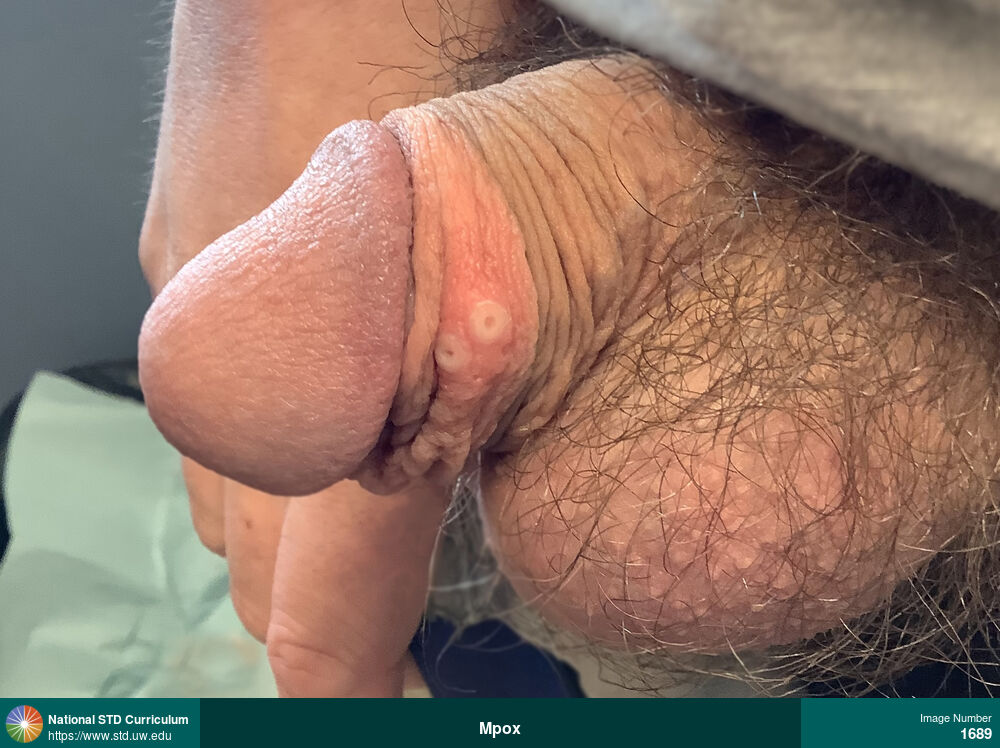
Mpox
Two round papular-pustular lesions with central crater on shaft of penis in a man with mpox.
Photo: Annular, Papule / Papules, Pustule / Pustules, Painful, Rash
Courtesy of Negusse Ocbamichael, PA
Annular, Papule / Papules, Pustule / Pustules N/A
1689
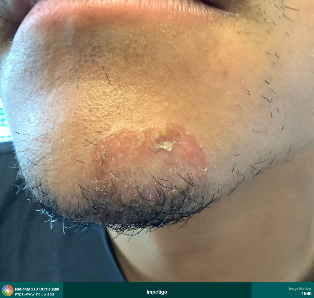
Impetigo
Large, round, erythematous, superficial ulcerated region on the lower left chin with yellow crusting in a man with impetigo. Several days prior, the affected area was a large bullae.
Photo: Bulla / Bullae, Erythema, Face, Rash
Courtesy of Negusse Ocbamichael, PA
Bulla / Bullae, Erythema Face
1690
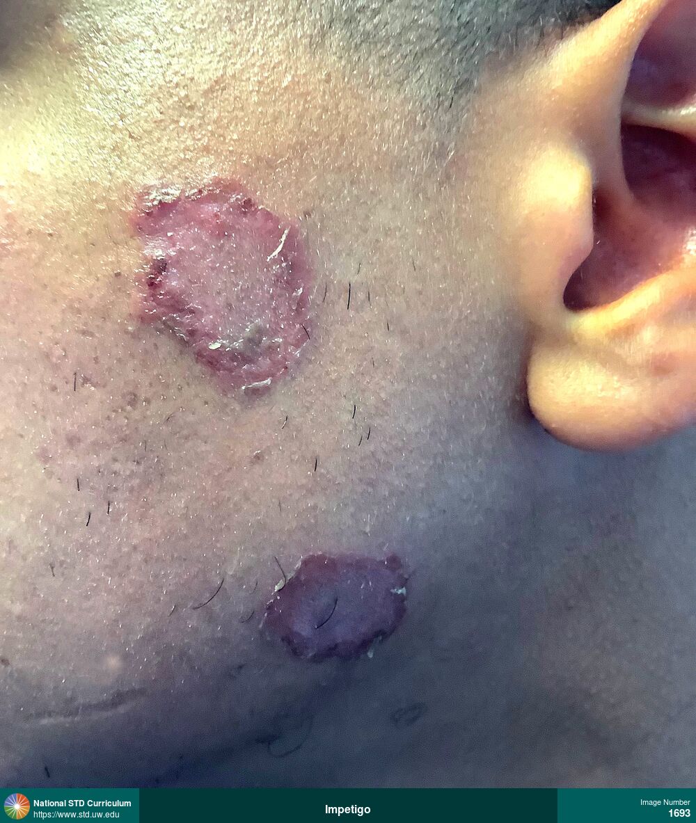
Impetigo
Two large, dark red, oval, superficial ulcerated regions (with crusting/scabbing) on the left side of the face in a man with impetigo. Several days prior, the affected areas were a large bullae.
Photo: Erythema, Hyperpigmentation, Patch/Patches, Scab, Face, Rash
Courtesy of Negusse Ocbamichael, PA
Erythema, Hyperpigmentation, Patch/Patches, Scab Face
1693
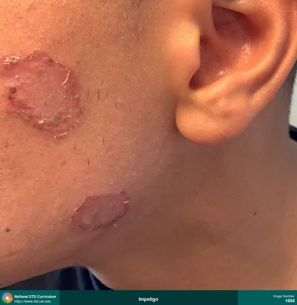
Impetigo
Two large, dark red, oval, superficial ulcerated regions (with crusting/scabbing) on the left side of the face in a man with impetigo. Several days prior, the affected areas were a large bullae.
Photo: Erythema, Hypopigmentation, Patch/Patches, Scab, Face, Rash
Courtesy of Negusse Ocbamichael, PA
Erythema, Hypopigmentation, Patch/Patches, Scab Face
1694
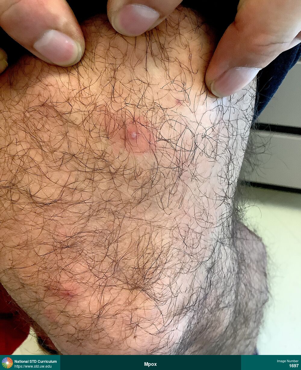
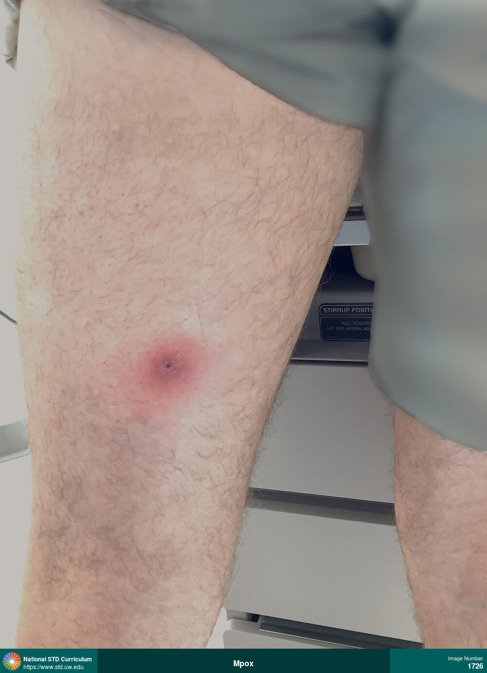
Mpox
Pustular mpox lesions (with surrounding erythema) on the left thigh.
Photo: Papule / Papules, Pustule / Pustules, Light skin tone, Thigh
Courtesy of Negusse Ocbamichael, PA
Papule / Papules, Pustule / Pustules Light skin tone, Thigh
1697
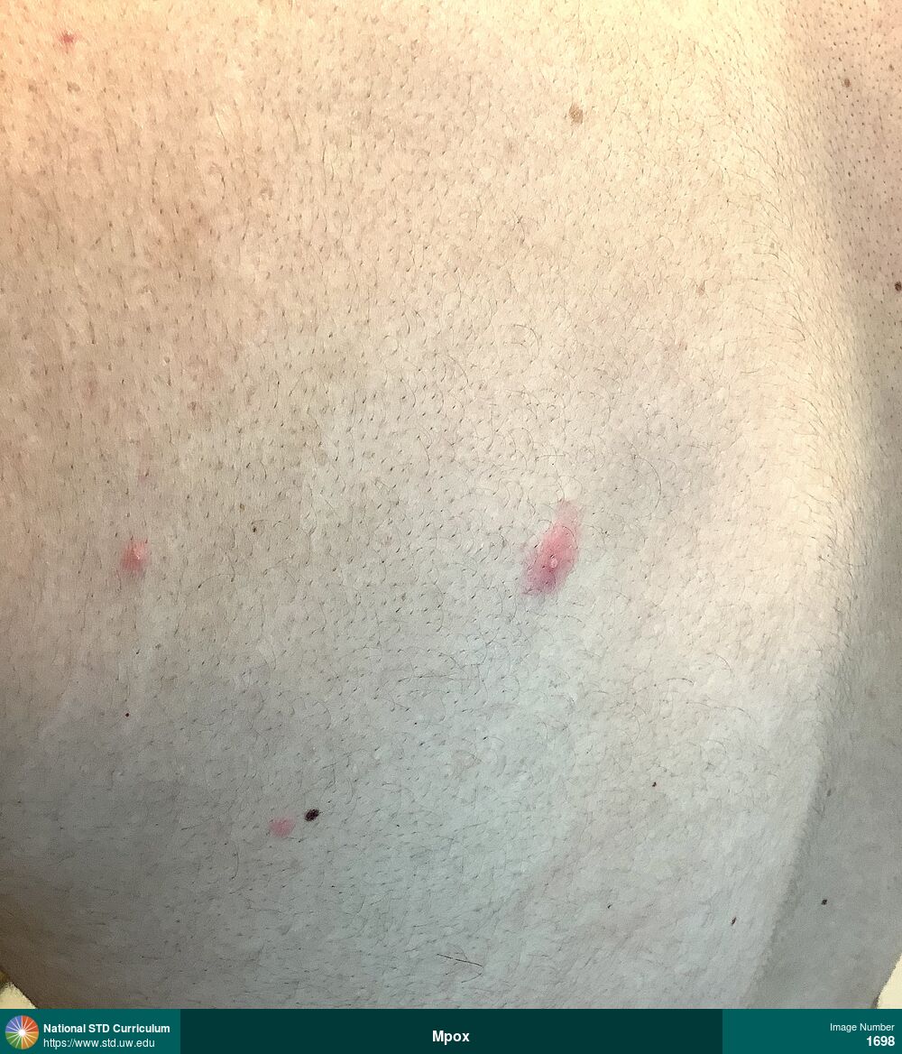
Mpox
Pustular mpox lesions (with surrounding erythema) on the back.
Photo: Papule / Papules, Pustule / Pustules, Back, Light skin tone, Non-Painful
Courtesy of Negusse Ocbamichael, PA
Papule / Papules, Pustule / Pustules Back, Light skin tone
1698
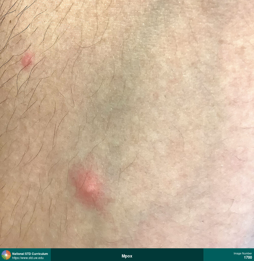
Mpox
Papular mpox lesions (with surrounding erythema) on the back.
Photo: Papule / Papules, Pustule / Pustules, Arm (Right), Light skin tone, Painful
Courtesy of Negusse Ocbamichael, PA
Papule / Papules, Pustule / Pustules Arm (Right), Light skin tone
1700

Mpox
Multiple mpox lesions on the penis, including lesions in pustular stage, ulcerative, and scabbing phase. There is severe edema of the penis.
Photo: Edema / Swelling, Pustule / Pustules, Ulcer / Ulcers, Dark skin tone, Penis, Painful
Courtesy of Negusse Ocbamichael, PA
Edema / Swelling, Pustule / Pustules, Ulcer / Ulcers Dark skin tone, Penis
1712
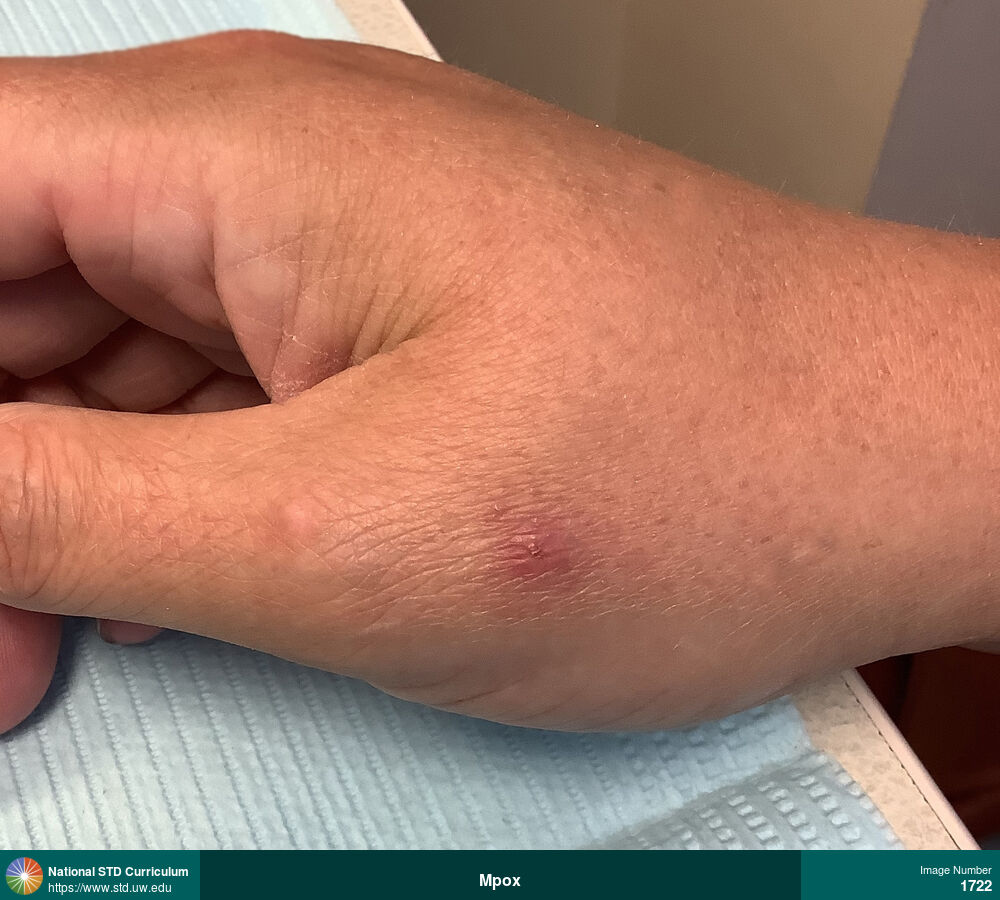
Mpox
Single, erythemaous papule at the base of the right thumb caused by mpox infection.
Photo: Erythema, Papule / Papules, Scab, Hand (Left), Painful
Courtesy of Negusse Ocbamichael, PA
Erythema, Papule / Papules, Scab Hand (Left)
1722
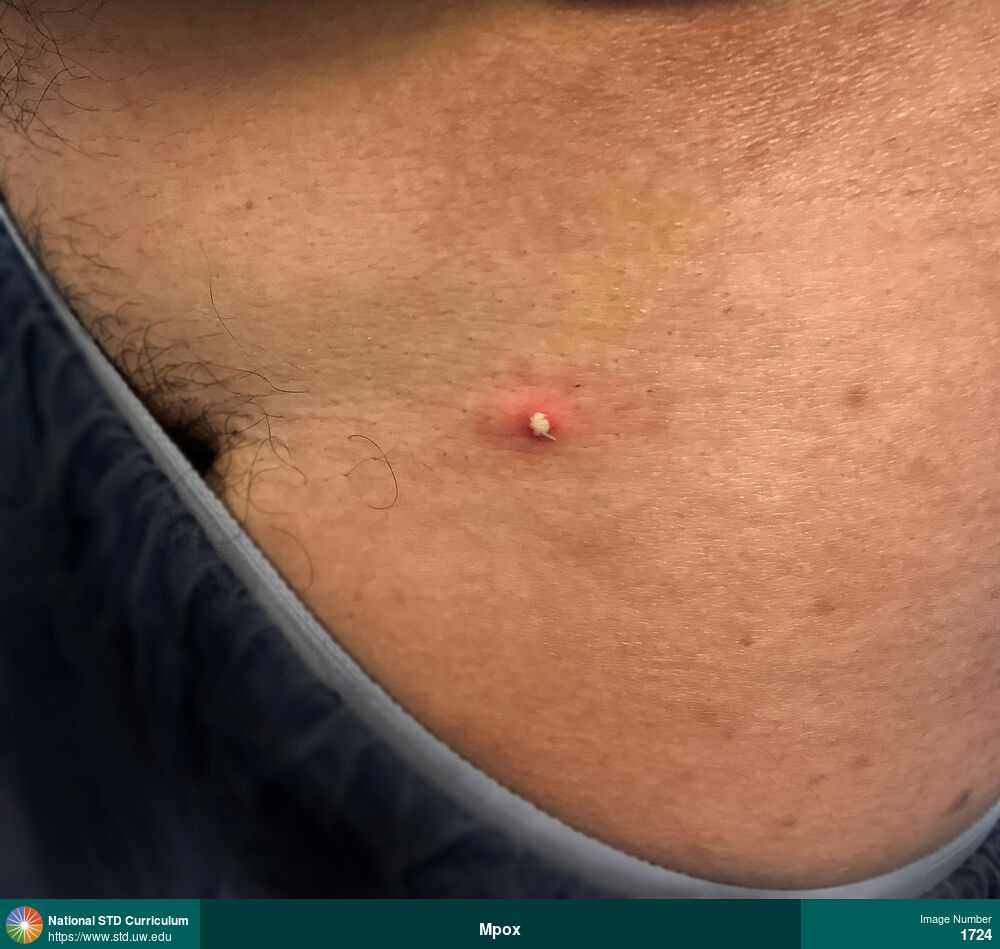
Mpox
Mpox pustular lesion on lower right back.
Photo: Erythema, Pustule / Pustules, Back
Courtesy of Negusse Ocbamichael, PA
Erythema, Pustule / Pustules Back
1724
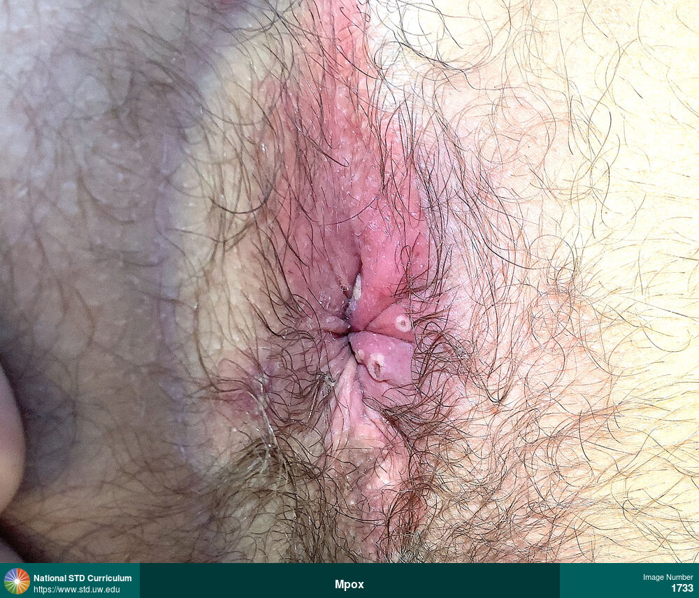
Mpox
Multiple painful, pustular, ulcerated anal and perianal lesions caused by mpox. Two of the anal lesions have a dark center.
Photo: Pustule / Pustules, Ulcer / Ulcers, Anal, Anal / Perianal, Painful
Courtesy of Negusse Ocbamichael, PA
Pustule / Pustules, Ulcer / Ulcers Anal, Anal / Perianal
1733
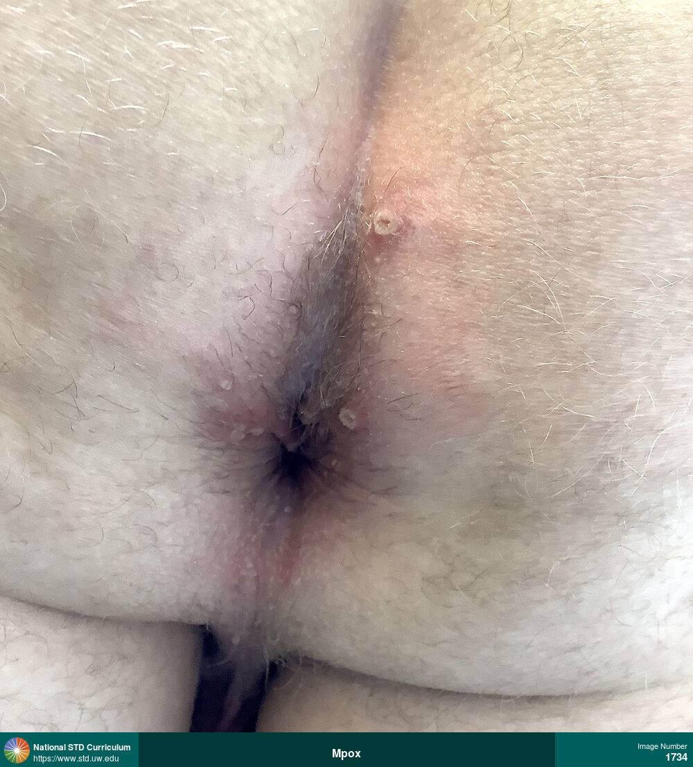
Mpox
Multiple painful anal and perianal vesicicular and pustular mpox lesions.
Photo: Erythema, Pustule / Pustules, Vesicle / Vesicles, Anal, Anal / Perianal, Buttock, Light skin tone, Painful, Rash
Courtesy of Negusse Ocbamichael, PA
Erythema, Pustule / Pustules, Vesicle / Vesicles Anal, Anal / Perianal, Buttock, Light skin tone
1734
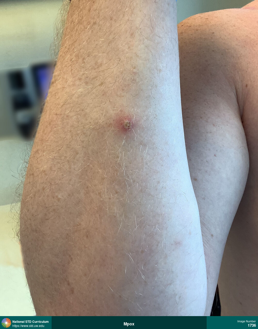
Mpox
Mpox pustule with surrounding erythema (right forearm).
Photo: Erythema, Pustule / Pustules, Arm (Right)
Courtesy of Negusse Ocbamichael, PA
Erythema, Pustule / Pustules Arm (Right)
1736
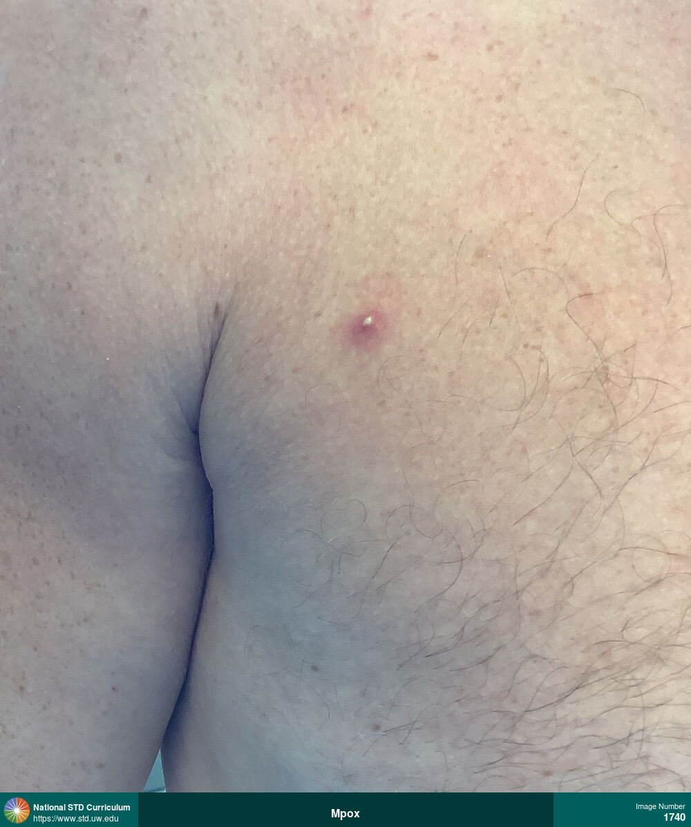
Mpox
Mpox pustule with surrounding erythema (right anterior chest region).
Photo: Erythema, Pustule / Pustules, Chest, Shoulder (Right)
Courtesy of Negusse Ocbamichael, PA
Erythema, Pustule / Pustules Chest, Shoulder (Right)
1740
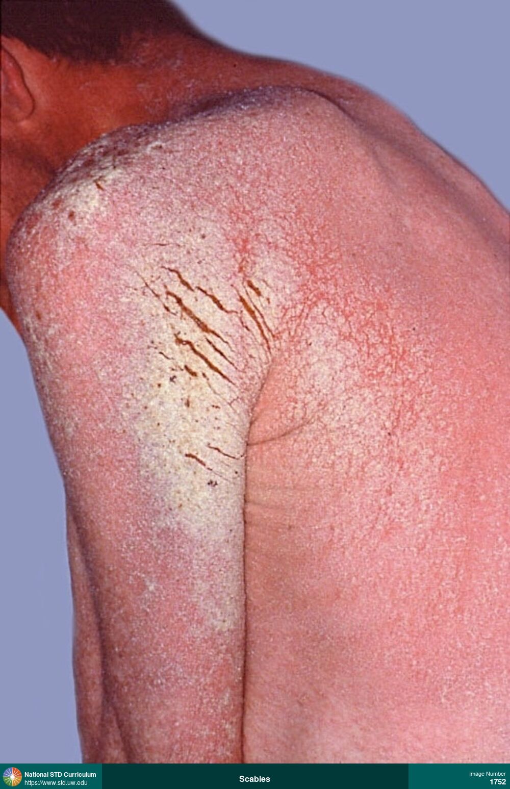
Scabies
Extensive plaque-like lesions on left shoulder caused by crusted scabies in a man with HIV and AIDS.
Photo: Plaque, Rash, Scale, Back, Shoulder (Left), Shoulder (Right), Non-Itchy, Rash
Courtesy of David H. Spach, MD
Plaque, Rash, Scale Back, Shoulder (Left), Shoulder (Right)
1752
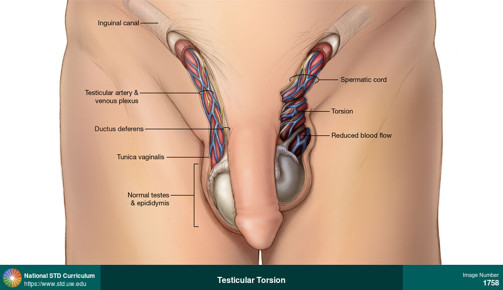
Testicular Torsion
This illustration of a young man with testicular torsion shows apparent twisting of the spermatic cord and reduced blood flow to the left testicle. The left testicle appears signifanctly elevated in the scrotum compared to the right testicle.
Illustration: Edema / Swelling, Epididymis, Light skin tone, Scrotum, Painful
Courtesy of Cognition Studio, Inc. and David H. Spach, MD
Edema / Swelling Epididymis, Light skin tone, Scrotum
1758
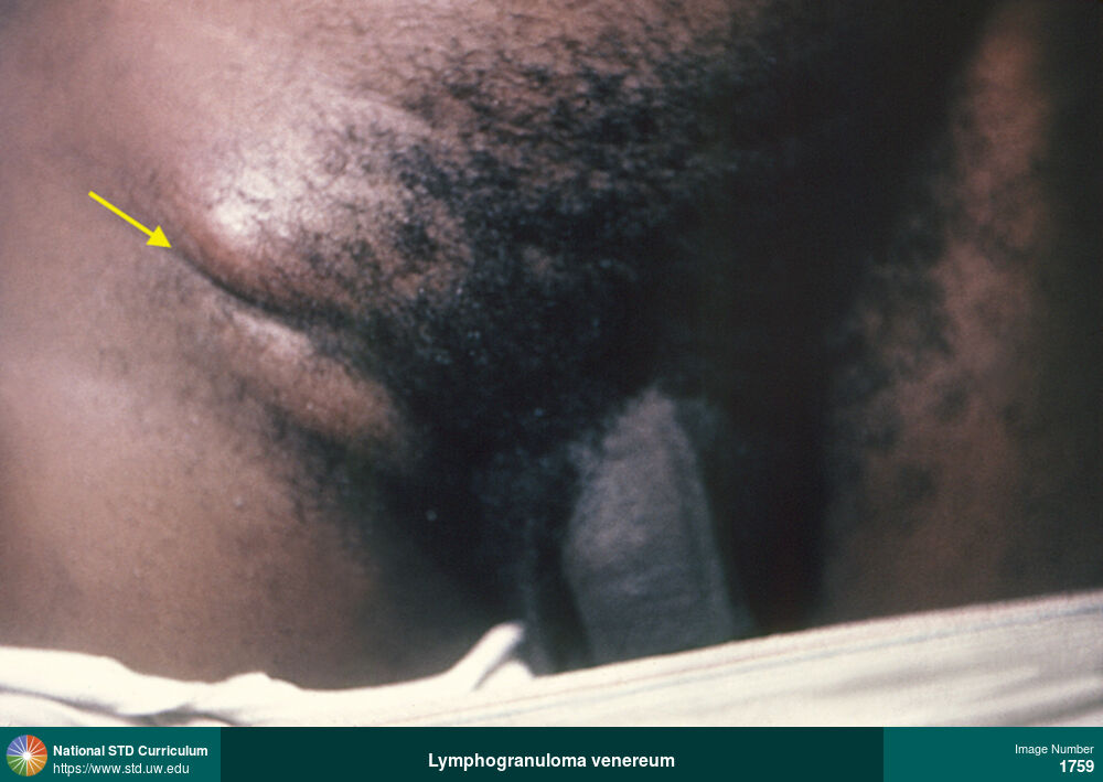
Lymphogranuloma venereum
Right-sided inguinal adenopathy “groove sign” as a manifestation of lymphogranuloma venereum (LGV). The groove sign is formed by a depression in enlarged inguinal lymph nodes that surround the inguinal ligament. Lymphogranuloma venereum is caused by L1, L2, or L3 serovars of Chlamydia trachomatis.
Photo: Edema / Swelling, Dark skin tone, Groin/Inguinal
Courtesy of CDC Public Health Image Library (Renelle Woodall)
Edema / Swelling Dark skin tone, Groin/Inguinal
1759
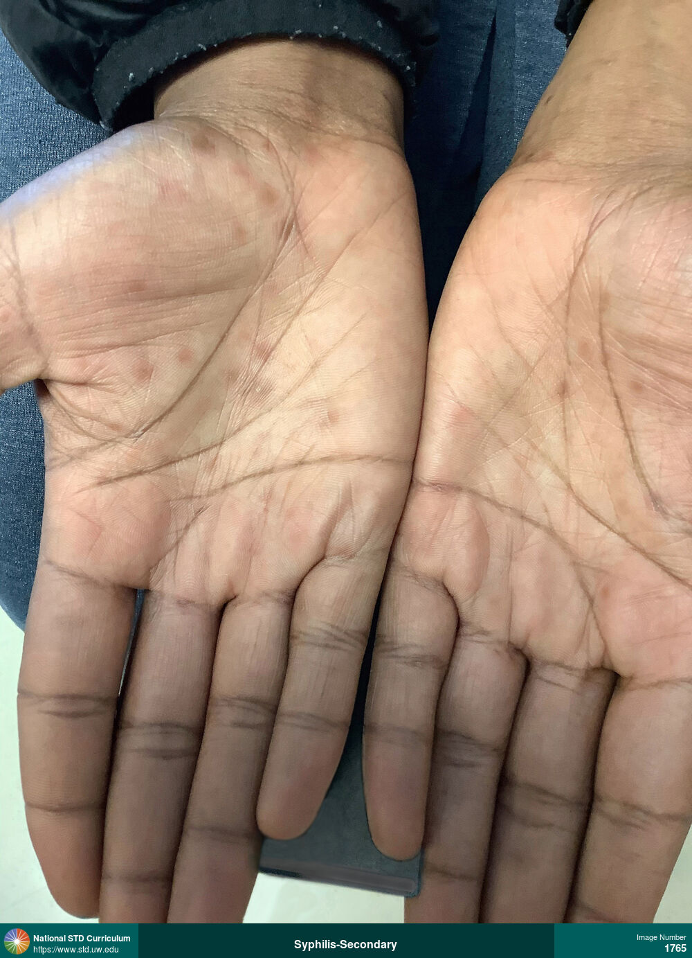
Syphilis-Secondary
Hyperpigmented macules on the palms in a woman with secondary syphilis.
Photo: Hyperpigmentation, Macule / Macules, Dark skin tone, Hand (Left), Hand (Right), Rash
Courtesy of Negusse Ocbamichael, PA
Hyperpigmentation, Macule / Macules Dark skin tone, Hand (Left), Hand (Right)
1765
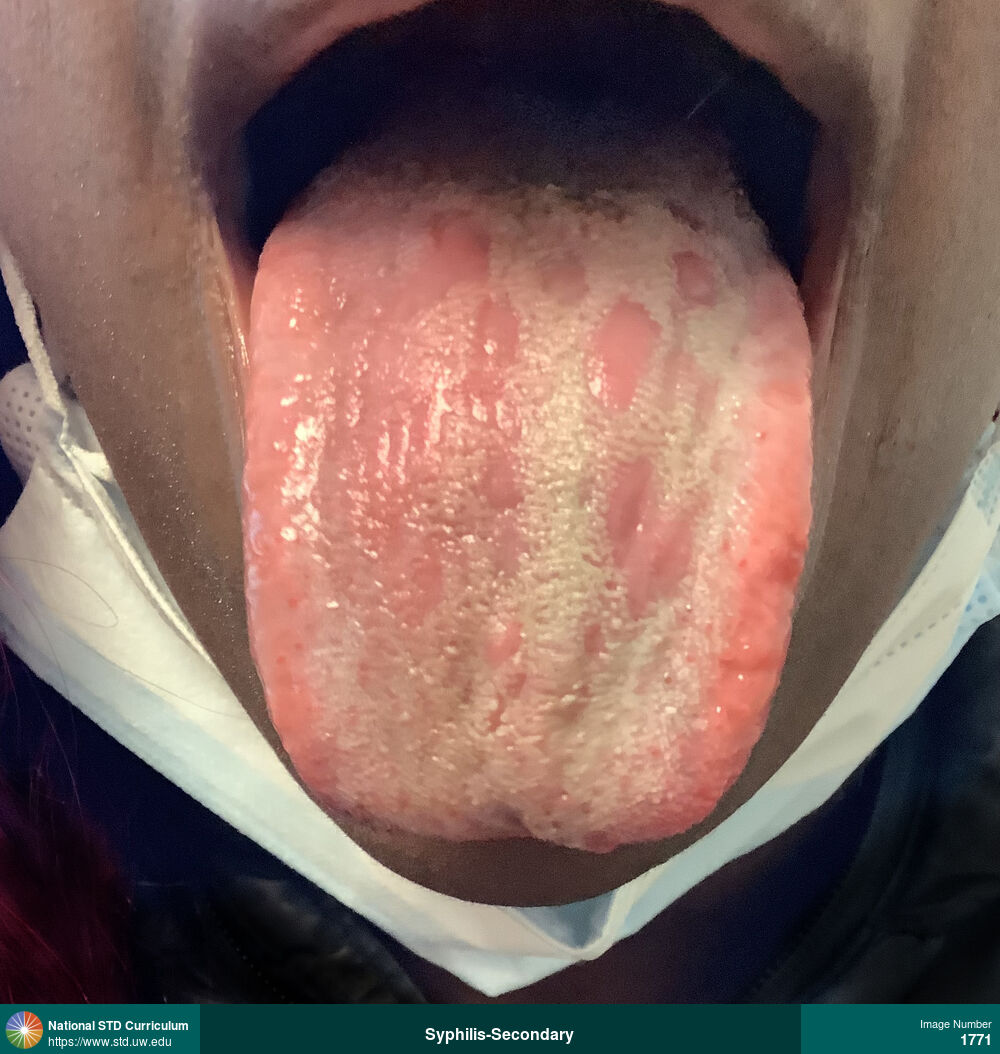
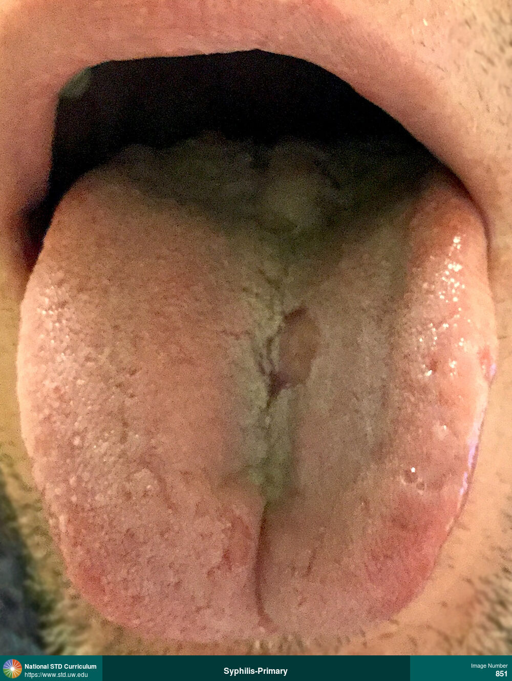
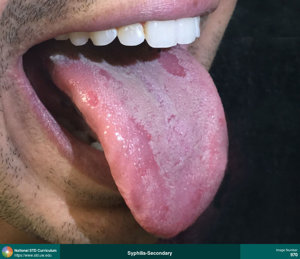
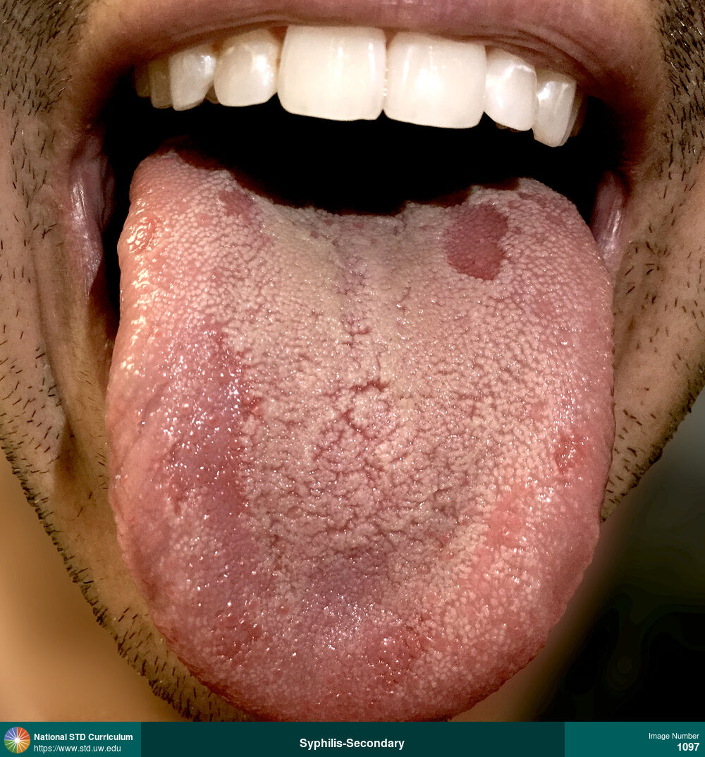
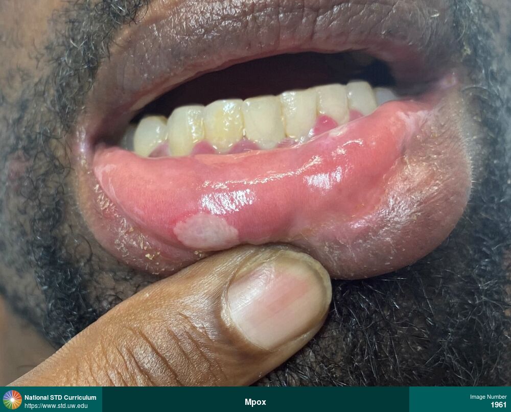
Syphilis-Secondary
Oral mucous patches as shown by superficial ulcerations on the tongue in a woman with secondary syphilis.
Photo: Patch/Patches, Ulcer / Ulcers, Dark skin tone, Tongue
Courtesy of Negusse Ocbamichael, PA
Patch/Patches, Ulcer / Ulcers Dark skin tone, Tongue
1771
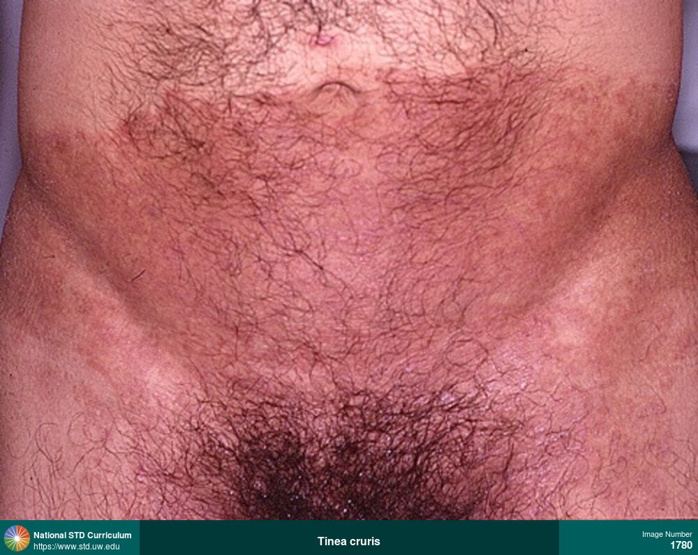
Tinea cruris
Tinea cruris in a man with HIV manifesting as an extensive, expanding, erythematous rash with a well-demarcated border involving in the groin, suprapubic region, and lower abdomen.
Photo: Erythema, Hyperpigmentation, Rash, Groin/Inguinal, Light skin tone, Suprapubic (Hypogastrium)
Courtesy of David H. Spach, MD
Erythema, Hyperpigmentation, Rash Groin/Inguinal, Light skin tone, Suprapubic (Hypogastrium)
1780
Fixed Drug Eruption
Fixed-drug ulcerated lesion rash on the tongue caused by trimethoprim-sulfamethoxazole.
Photo: Annular, Ulcer / Ulcers, Dark skin tone, Tongue
Courtesy of Negusse Ocbamichael, PA
Annular, Ulcer / Ulcers Dark skin tone, Tongue
1795
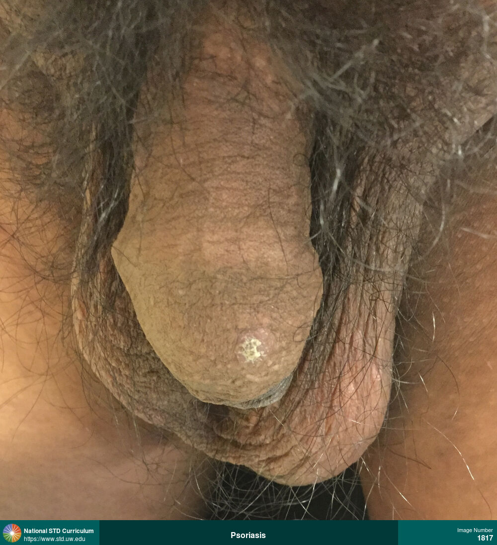
Psoriasis
Psoriasis plaque on glans penis in a man that has recurrent psoriasis on his hands and penis.
Photo: Papule / Papules, Plaque, Scale, Dark skin tone, Penis, Non-Painful
Courtesy of Negusse Ocbamichael, PA
Papule / Papules, Plaque, Scale Dark skin tone, Penis
1817
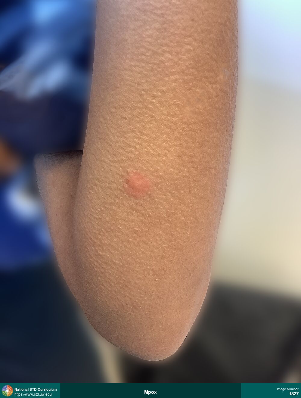
Mpox
Erythematous papular lesion on left forearm caused by mpox.
Photo: Erythema, Papule / Papules, Arm (Left)
Courtesy of Negusse Ocbamichael, PA
Erythema, Papule / Papules Arm (Left)
1827
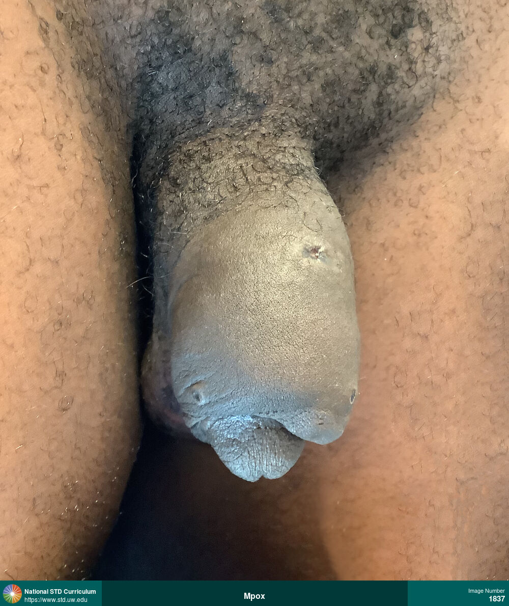
Mpox
Multiple mpox lesions, at various phases, on the penis and associated severe edema of the penis.
Photo: Edema / Swelling, Pustule / Pustules, Scab, Ulcer / Ulcers, Dark skin tone, Penis, Painful
Courtesy of Negusse Ocbamichael, PA
Edema / Swelling, Pustule / Pustules, Scab, Ulcer / Ulcers Dark skin tone, Penis
1837
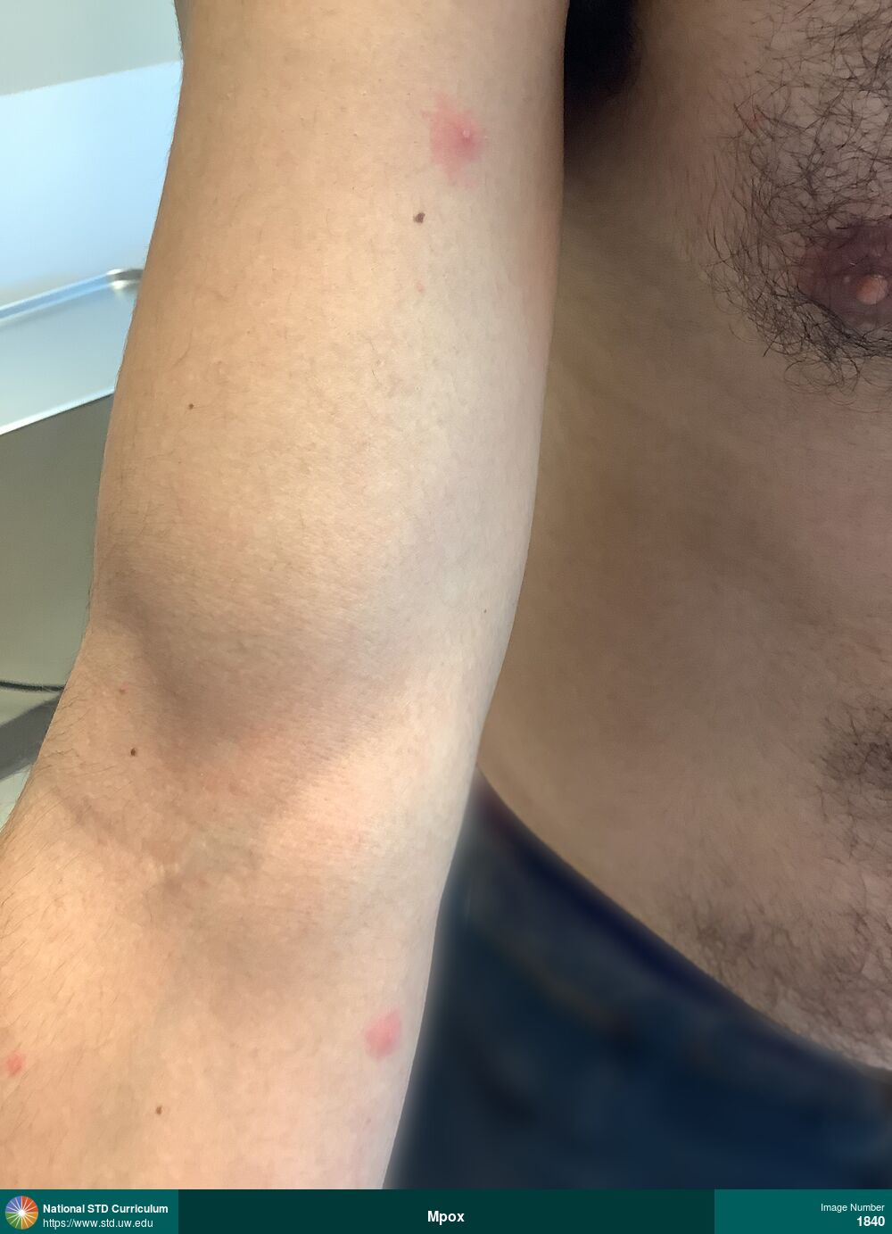
Mpox
Pustular mpox lesions (with surrounding erythema) on the right arm.
Photo: Papule / Papules, Pustule / Pustules, Arm (Right), Non-Painful
Courtesy of Negusse Ocbamichael, PA
Papule / Papules, Pustule / Pustules Arm (Right)
1840
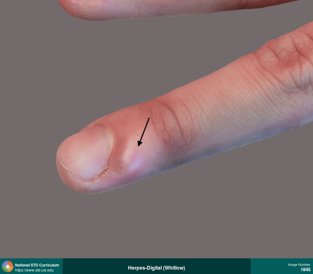
Herpes-Digital (Whitlow), Herpes-Genital
Photo: Vesicle / Vesicles, Hand (Left), Light skin tone, Non-Itchy, Painful
Courtesy of Negusse Ocbamichael, PA
Vesicle / Vesicles Hand (Left), Light skin tone
1845
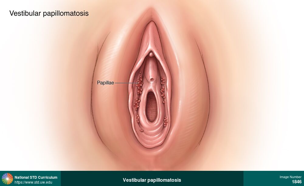
Vestibular papillomatosis
Vestibular papillomatosis is a normal anatomic variation characterized by multiple, small, smooth, finger-like (filiform) projections that symmetrically line the inner surface of the labia minora.
Illustration: Papule / Papules, Labia (majora/minora), Light skin tone, Non-Itchy, Non-Painful
Courtesy of Cognition Studio, Inc. and David H. Spach, MD
Papule / Papules Labia (majora/minora), Light skin tone
1846
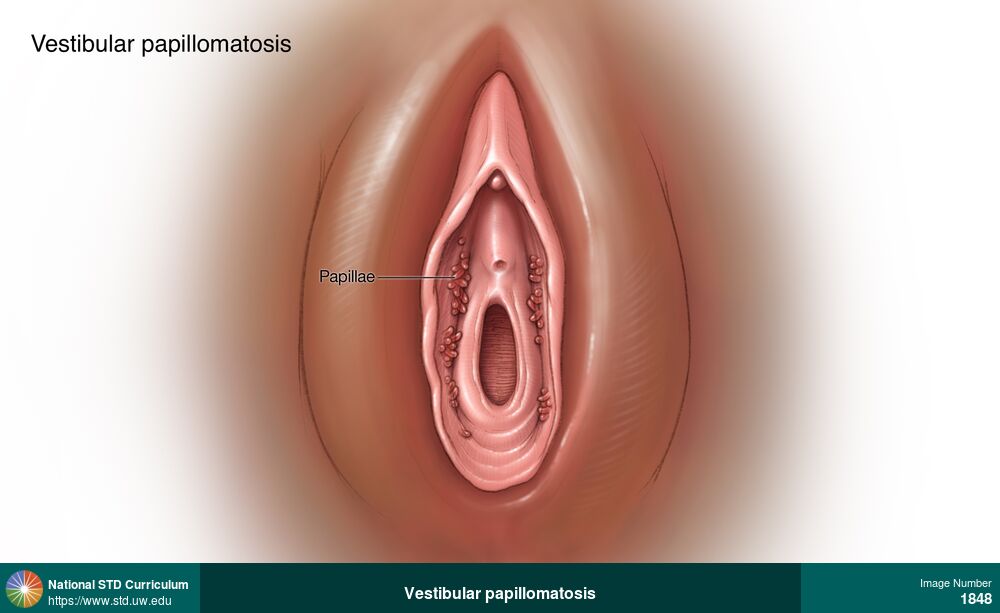
Vestibular papillomatosis
Vestibular papillomatosis is a normal anatomic variation characterized by multiple, small, smooth, finger-like (filiform) projections that symmetrically line the inner surface of the labia minora.
Illustration: Papule / Papules, Dark skin tone, Labia (majora/minora), Non-Itchy, Non-Painful
Courtesy of Cognition Studio, Inc. and David H. Spach, MD
Papule / Papules Dark skin tone, Labia (majora/minora)
1848

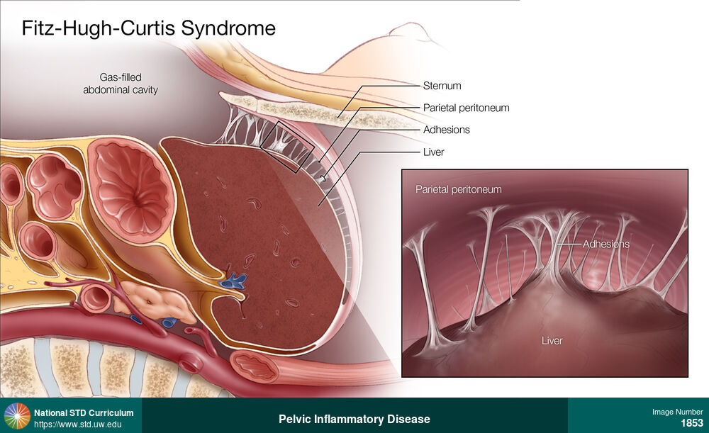
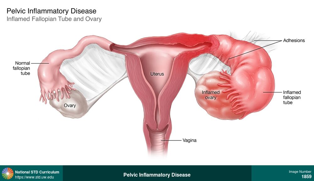
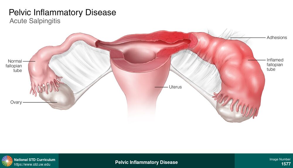
Pelvic Inflammatory Disease
This illustration of a woman with pelvic inflammatory disease shows inflammation and edema of the left fallopian tube and left ovary, with adhesions between the fallopian tube and the left ovary.
Illustration: Edema / Swelling, Female Genitourinary, Painful
Courtesy of Cognition Studio, Inc. and David H. Spach, MD
Edema / Swelling Female Genitourinary
1859
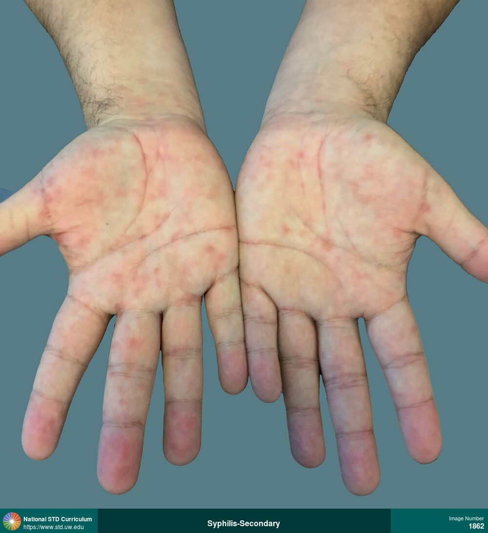
Syphilis-Secondary
Hyperpigmented macular lesions on palms in a man with secondary syphilis.
Photo: Erythema, Macule / Macules, Hand (Left), Hand (Right), Non-Itchy, Non-Painful, Rash
Courtesy of Negusse Ocbamichael, PA
Erythema, Macule / Macules Hand (Left), Hand (Right)
1862
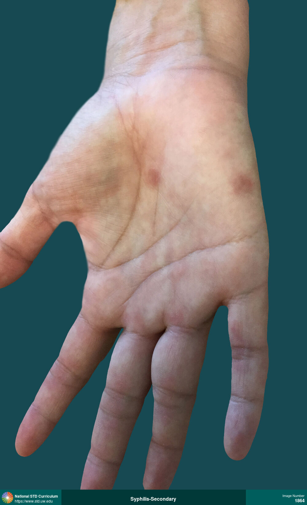
Syphilis-Secondary
Hyperpigmented maculopapular rash on right palm in a man with secondary syphilis.
Photo: Annular, Hyperpigmentation, Macule / Macules, Papule / Papules, Hand (Right), Non-Painful, Rash
Courtesy of Negusse Ocbamichael, PA
Annular, Hyperpigmentation, Macule / Macules, Papule / Papules Hand (Right)
1864
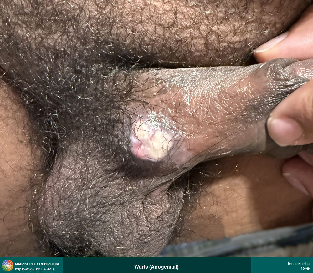
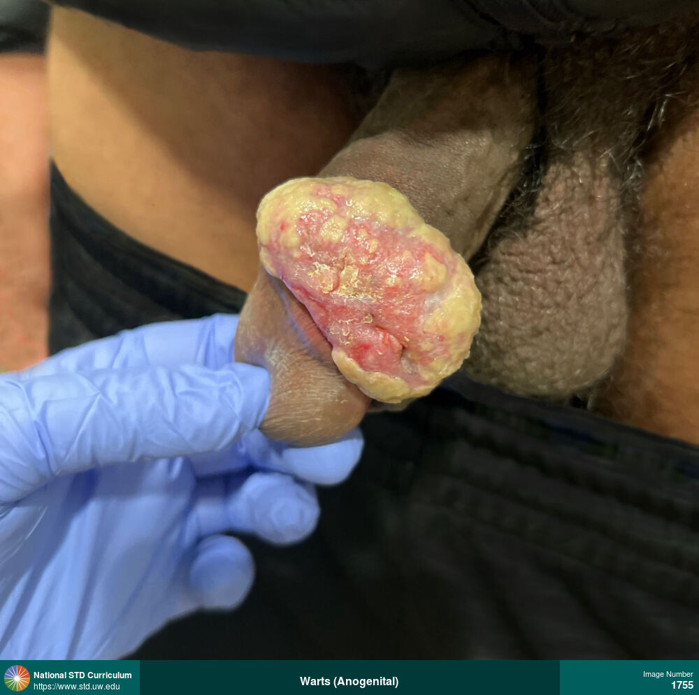
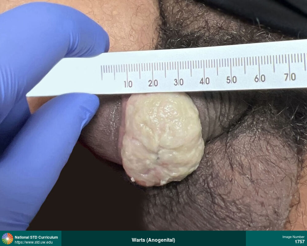
Warts (Anogenital)
Verrucous mass on the proximal shaft of the penis in a man with HIV. This mass was a recurrence of Buschke-Löwenstein tumor (also referred to as giant condyloma acuminata). The prior mass was located on the distal shaft of the penis and was surgically removed.
Photo: Mass, Verrucous, Dark skin tone, Penis
Courtesy of Olympia Stafford, MD
Mass, Verrucous Dark skin tone, Penis
1865

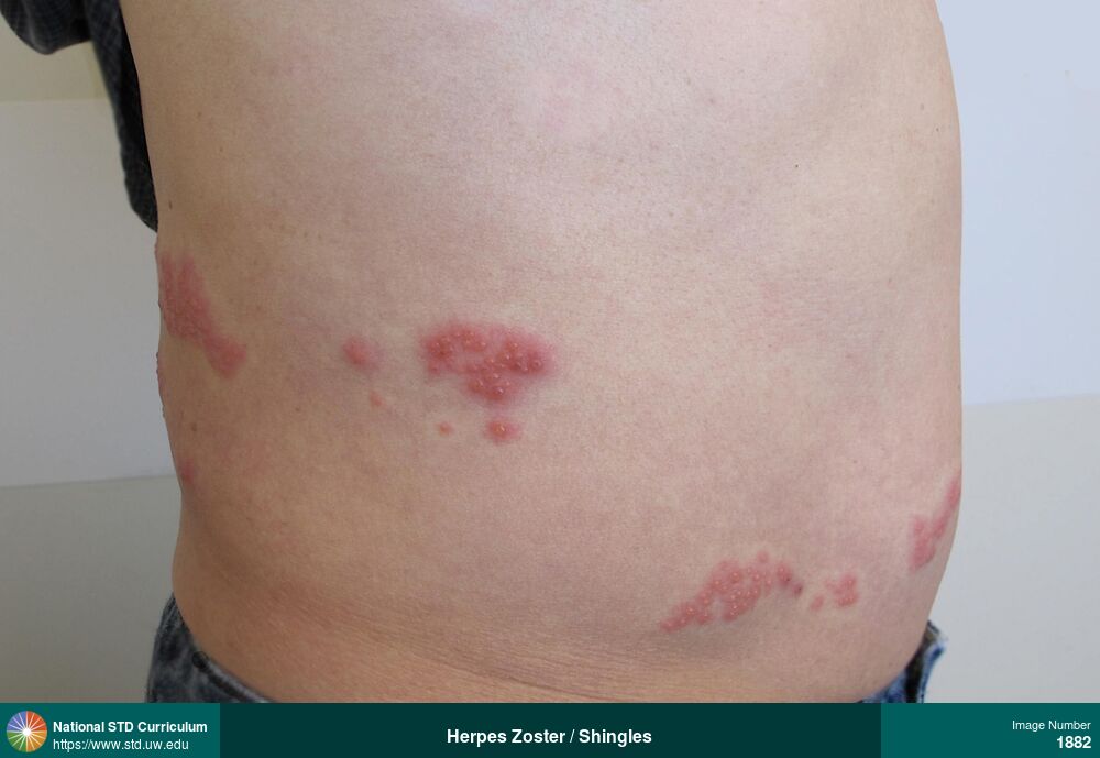
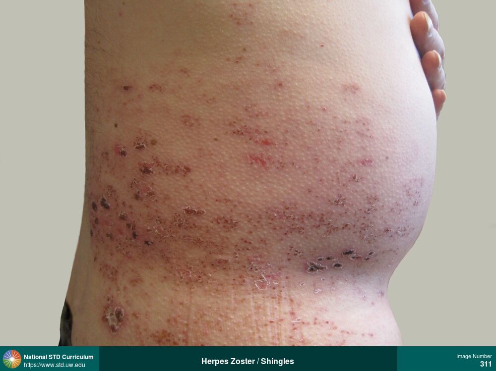
Herpes Zoster / Shingles
Photo: Erythema, Vesicle / Vesicles, Abdomen, Back, Light skin tone, Painful
Courtesy of Negusse Ocbamichael, PA
Erythema, Vesicle / Vesicles Abdomen, Back, Light skin tone
1882
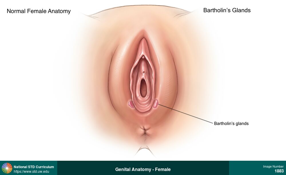
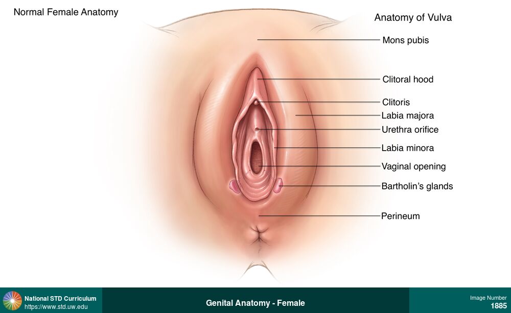
Genital Anatomy - Female
Bartholin’s glands are approximately 0.5 mm in size, oval-shaped, and located bilaterally in the lower region of the labia at the vaginal introitus. The duct of the Bartholin’s gland drains into the vaginal vestibule. These glands produce a clear mucous fluid that provides lubrication for the vagina and vulva.
Illustration: Labia (majora/minora), Light skin tone, Vulva
Courtesy of Cognition Studio, Inc. and David H. Spach, MD
N/A Labia (majora/minora), Light skin tone, Vulva
1883
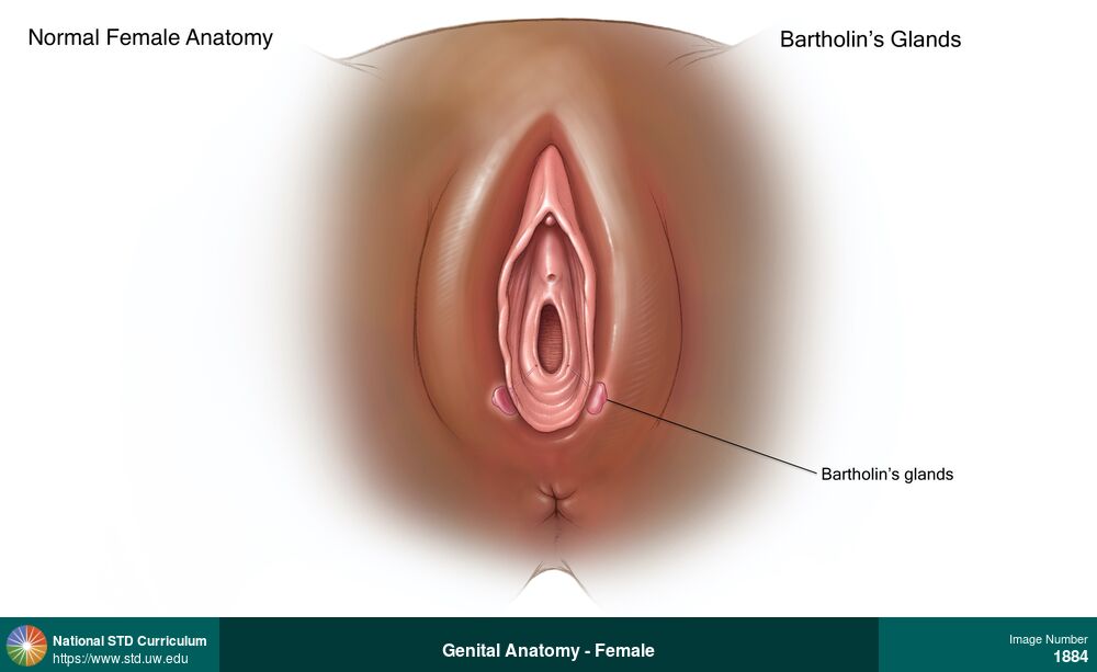
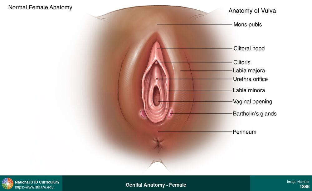
Genital Anatomy - Female
Bartholin’s glands are approximately 0.5 mm in size, oval-shaped, and located bilaterally in the lower region of the labia at the vaginal introitus. The duct of the Bartholin’s gland drains into the vaginal vestibule. These glands produce a clear mucous fluid that provides lubrication for the vagina and vulva.
Illustration: Dark skin tone, Labia (majora/minora), Vulva
Courtesy of Cognition Studio, Inc. and David H. Spach, MD
N/A Dark skin tone, Labia (majora/minora), Vulva
1884
Genital Anatomy - Female
External reproductive system (vulva) anatomy.
Illustration: Dark skin tone, Labia (majora/minora)
Courtesy of Cognition Studio, Inc. and David H. Spach, MD
N/A Dark skin tone, Labia (majora/minora)
1886
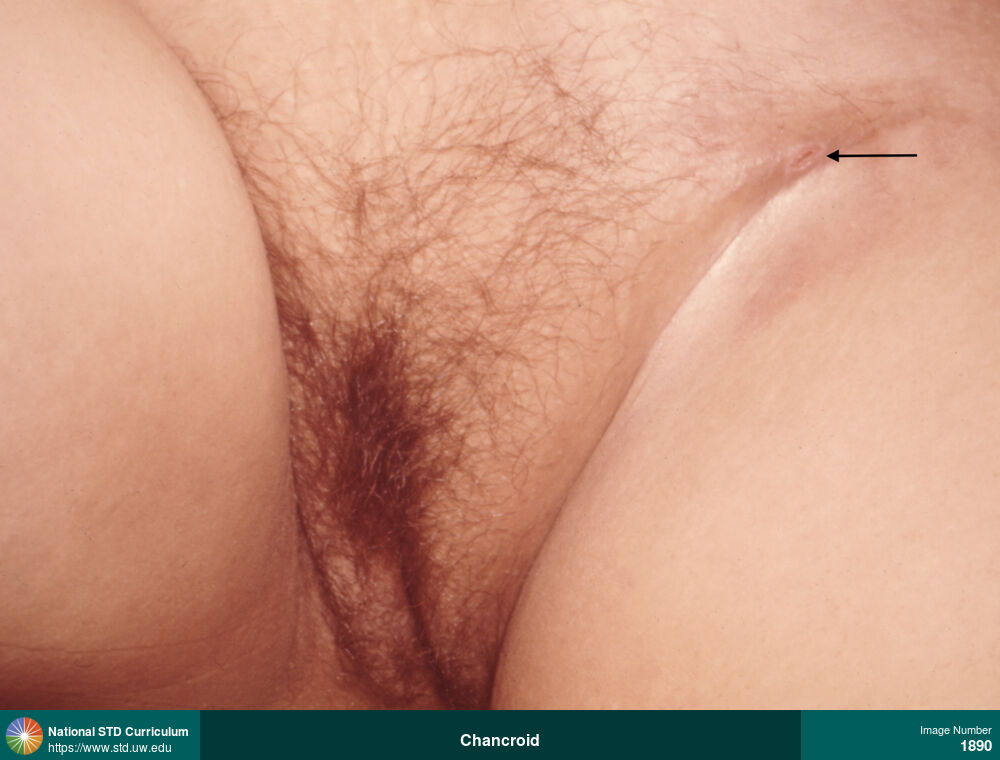
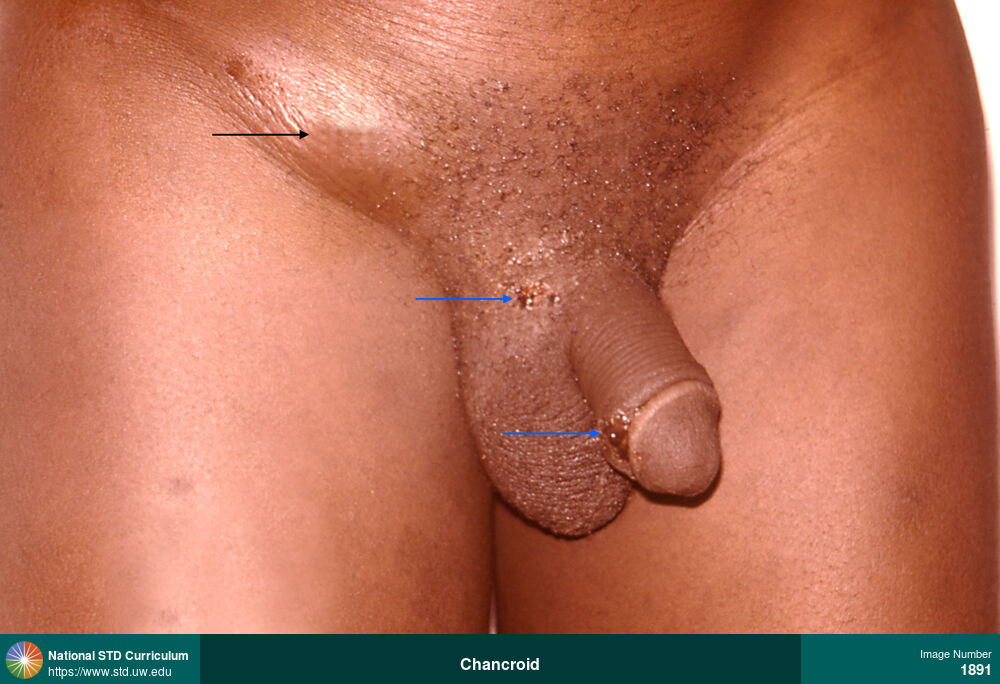
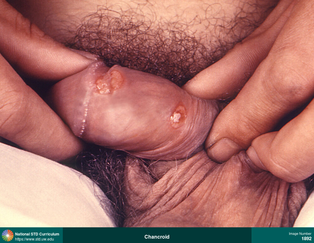
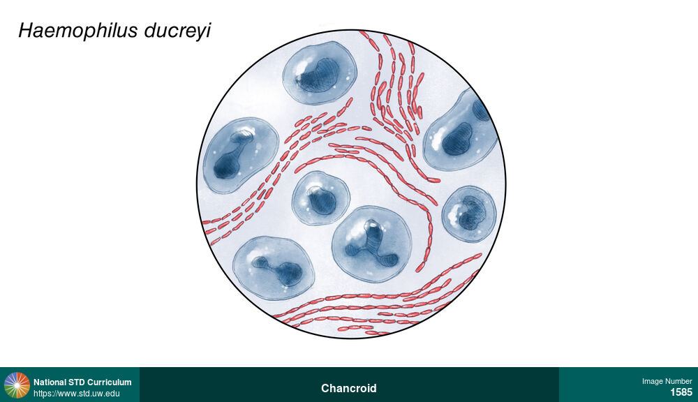
Chancroid
This photograph of a man with chancroid shows shallow, soft ulcers on the penis, covered with a yellow exudate. Chancroid is caused by the bacterium Haemophilus ducreyi.
Photo: Ulcer / Ulcers, Dark skin tone, Painful
Courtesy of CDC Public Health Image Library (Dr. Pirozzi)
Ulcer / Ulcers Dark skin tone
1892
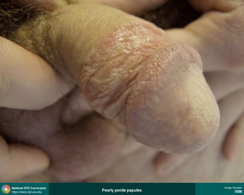
Pearly penile papules
Pearly penile papules are a normal anatomic variant that appear as small (<1 mm), flesh-colored bumps in rows around the corona of the penis.
Photo: Papule / Papules, Light skin tone, Penis, Non-Itchy, Non-Painful
Courtesy of Seattle & King County Sexual Health Clinic
Papule / Papules Light skin tone, Penis
1896

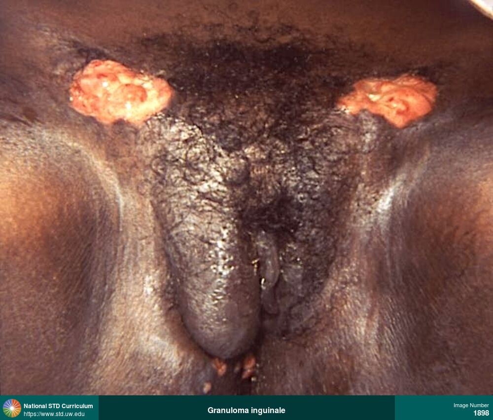
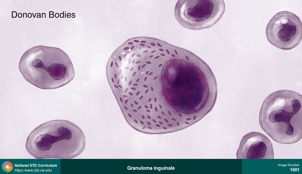
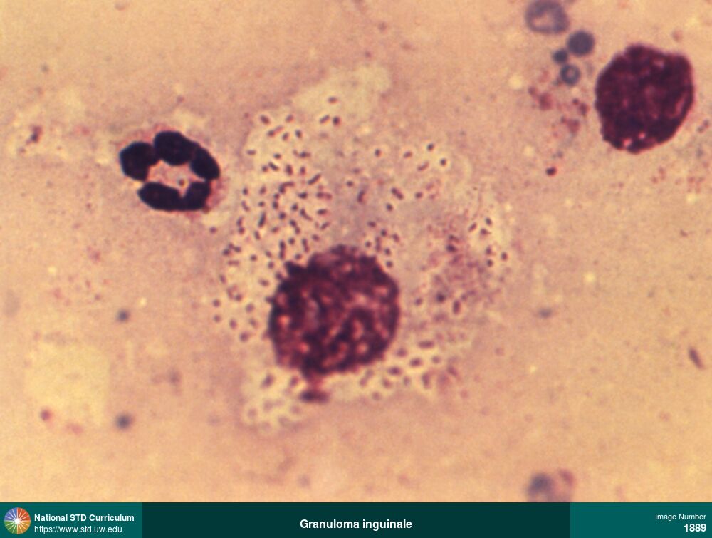
Granuloma inguinale
Large bilateral inguinal ulcers in a woman with granuloma inguinale (Donovanosis). The ulcers have progressed with raised borders, abundant central granulation tissue, and hypopigmentation. There is also significant edema in the right vulvar region. Granuloma inguinale is a sexually transmitted infection caused by the gram-negative bacteria Klebsiella granulomatis.
Photo: Hypopigmentation, Ulcer / Ulcers, Dark skin tone, Groin/Inguinal, Vulva, Non-Painful
Courtesy of CDC Public Health Image Library (Susan Lindsley)
Hypopigmentation, Ulcer / Ulcers Dark skin tone, Groin/Inguinal, Vulva
1898
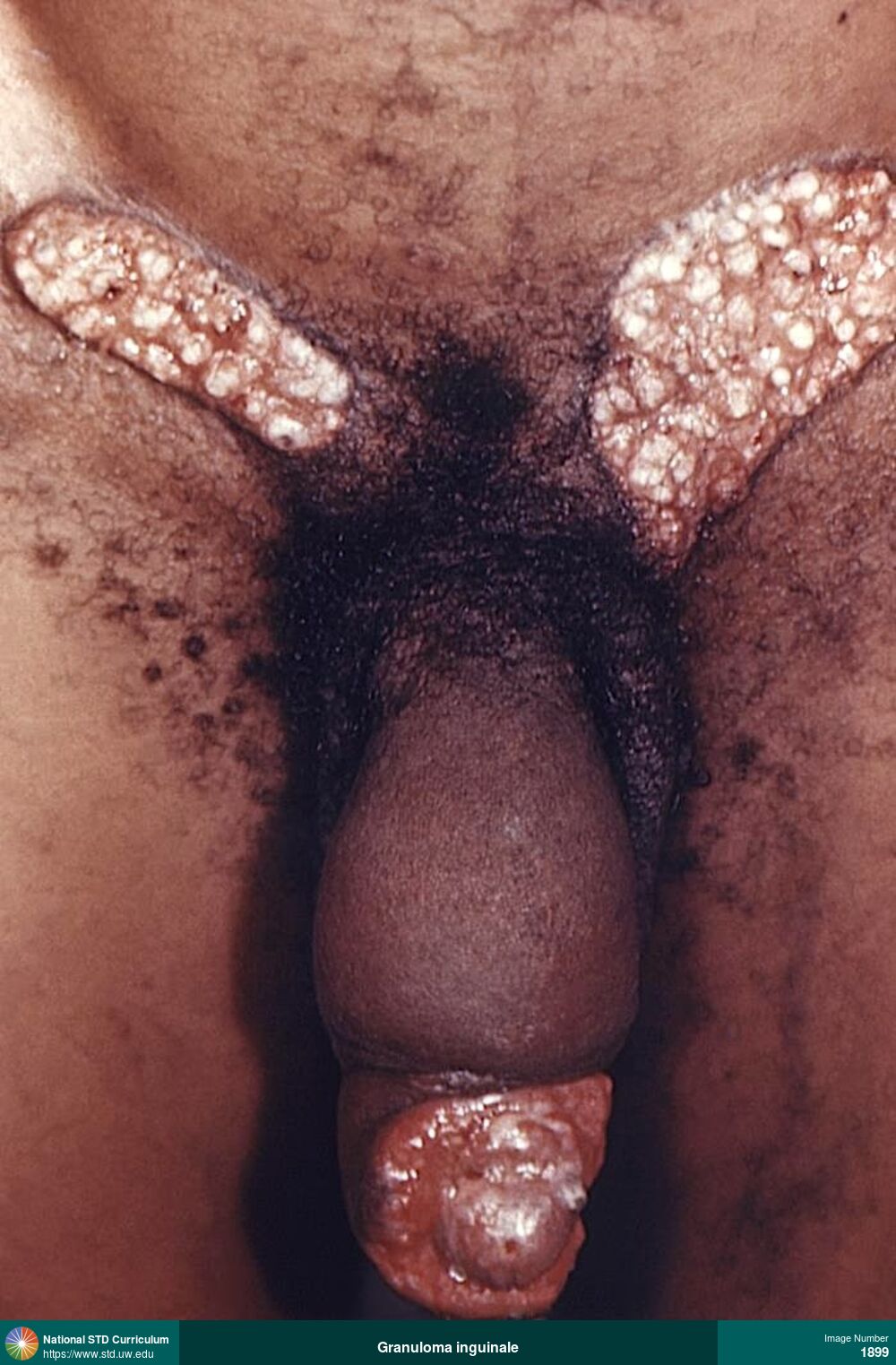
Granuloma inguinale
Large bilateral inguinal and penile ulcers in a man with granuloma inguinale (Donovanosis). There marked formation of raised hypopigmented granulation tissue in both inguinal ulcers. There is also extensive edema of the penis. Granuloma inguinale is a sexually transmitted infection caused by the gram-negative bacteria Klebsiella granulomatis.
Photo: Edema / Swelling, Dark skin tone, Groin/Inguinal, Penis
Courtesy of CDC Public Health Image Library (Renelle Woodall)
Edema / Swelling Dark skin tone, Groin/Inguinal, Penis
1899
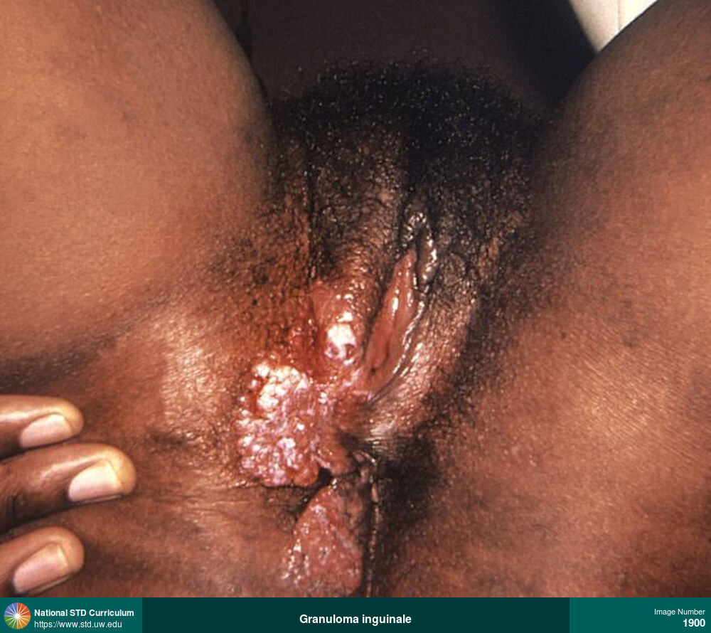
Granuloma inguinale
Large bilateral vulvar, perineal, and ulcers in a woman with granuloma inguinale (Donovanosis). The ulcers have progressed with raised borders, with abundant granulation tissue. Granuloma inguinale is a sexually transmitted infection caused by the gram-negative bacteria Klebsiella granulomatis.
Photo: Edema / Swelling, Ulcer / Ulcers, Anal / Perianal, Dark skin tone, Vulva
Courtesy of CDC Public Health Image Library (Dr. Peter Perine)
Edema / Swelling, Ulcer / Ulcers Anal / Perianal, Dark skin tone, Vulva
1900
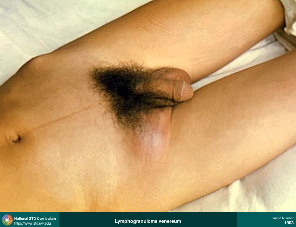
Lymphogranuloma venereum
Large, right-sided, fluctuant, inguinal lymph node (“bubo”) caused by lymphogranuloma venereum (LGV). There is erythema on the skin overlying the swollen lymph node. Lymphogranuloma venereum is caused by C. trachomatis serovars L1, L2, or L3.
Photo: Adenopathy, Edema / Swelling, Erythema, Groin/Inguinal
Courtesy of CDC Public Health Image Library (O.T. Chambers)
Adenopathy, Edema / Swelling, Erythema Groin/Inguinal
1903
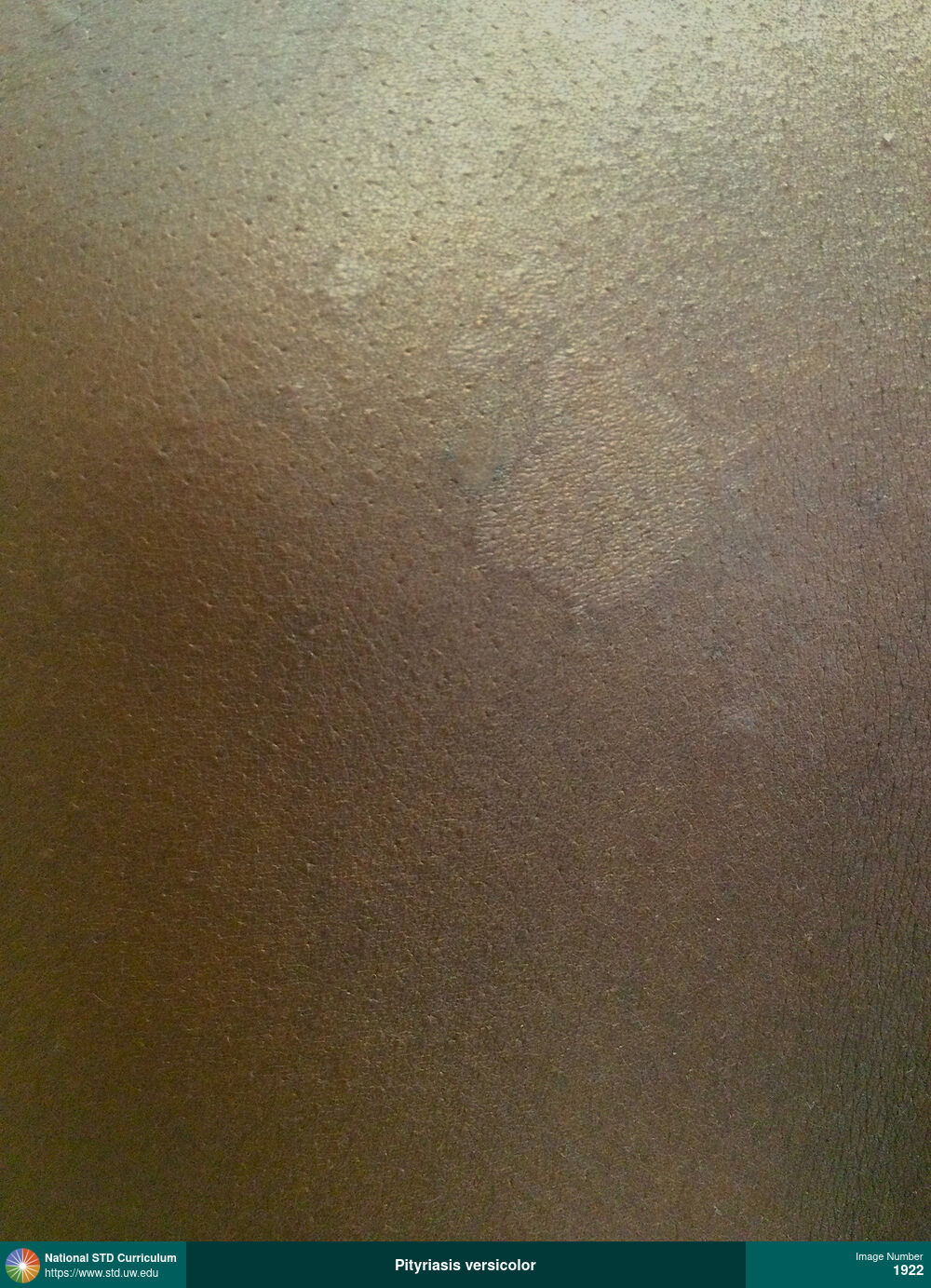
Pityriasis versicolor
Hypopigmented patches are present on the left upper back region. The diagnosis was pityriasis versicolor (also known as tinea versicolor). This condition is caused by overgrowth of a certain type of yeast (Malassezia) on the skin.
Photo: Hypopigmentation, Macule / Macules, Patch/Patches, Rash, Back, Non-Itchy, Non-Painful
Courtesy of Negusse Ocbamichael, PA
Hypopigmentation, Macule / Macules, Patch/Patches, Rash Back
1922
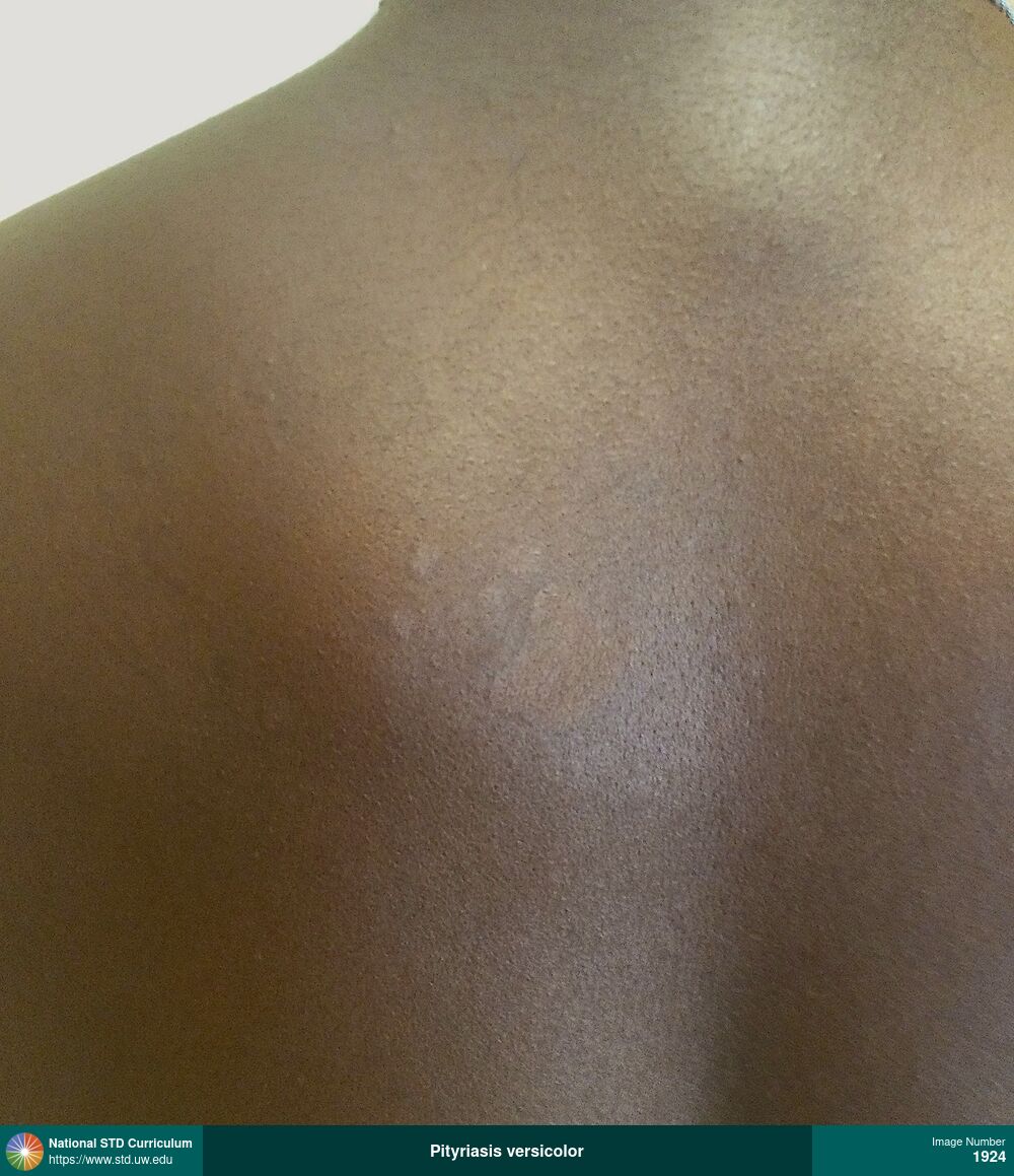
Pityriasis versicolor
Hypopigmented patches are present on the left upper back region. The diagnosis was pityriasis versicolor (also known as tinea versicolor). This condition is caused by overgrowth of a certain type of yeast (Malassezia) on the skin.
Photo: Hypopigmentation, Macule / Macules, Patch/Patches, Rash, Back, Dark skin tone, Non-Itchy, Non-Painful
Courtesy of Negusse Ocbamichael, PA
Hypopigmentation, Macule / Macules, Patch/Patches, Rash Back, Dark skin tone
1924
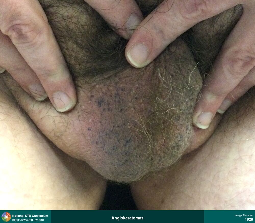
Angiokeratomas
Multiple, purple, maculo-papular lesions on the scrotum caused angiokeratomas, which are caused by benign capillary ectasias.
Photo: Hyperpigmentation, Macule / Macules, Papule / Papules, Light skin tone, Scrotum, Non-Itchy, Non-Painful
Courtesy of Negusse Ocbamichael, PA
Hyperpigmentation, Macule / Macules, Papule / Papules Light skin tone, Scrotum
1928
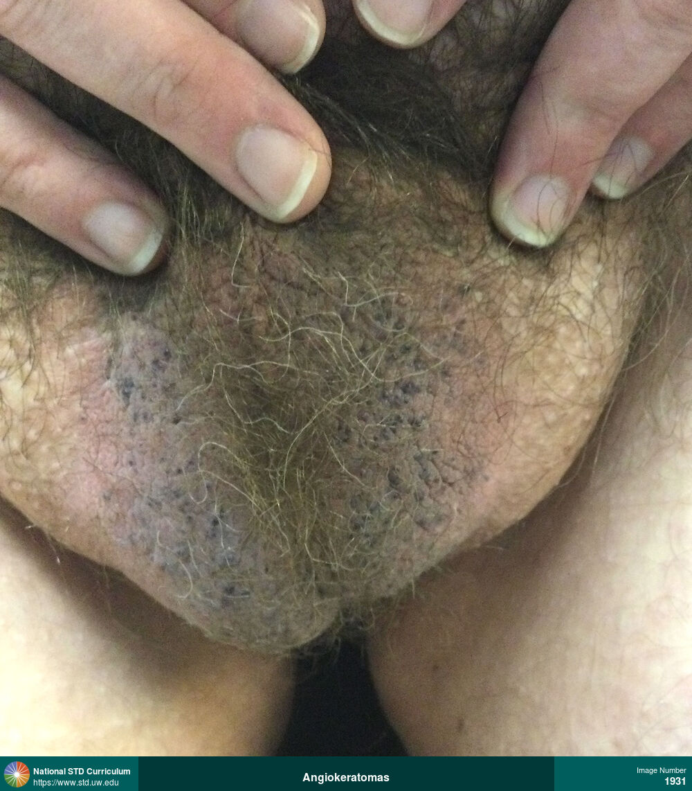
Angiokeratomas
Multiple, purple, maculo-papular lesions on the scrotum caused angiokeratomas, which are caused by benign capillary ectasias.
Photo: Hyperpigmentation, Macule / Macules, Papule / Papules, Light skin tone, Scrotum, Non-Itchy, Non-Painful, Rash
Courtesy of Negusse Ocbamichael, PA
Hyperpigmentation, Macule / Macules, Papule / Papules Light skin tone, Scrotum
1931

Lichen simplex chronicus
The man presented with pruritic, scaling, excoriated plaques over the scrotum and upper inner thighs bilaterally. These lesions were caused by chronic scratching. The diagnosis was lichen simplex chronicus, which is also known as neurodermatitis.
Photo: Hyperpigmentation, Rash, Scale, Dark skin tone, Groin/Inguinal, Scrotum, Itch
Courtesy of Negusse Ocbamichael, PA
Hyperpigmentation, Rash, Scale Dark skin tone, Groin/Inguinal, Scrotum
1932

Epididymitis
Marked left testicular and epididymal swelling and erythema caused by epididymitis.
Photo: Edema / Swelling, Erythema, Epididymis, Light skin tone, Scrotum, Painful
Courtesy of Negusse Ocbamichael, PA
Edema / Swelling, Erythema Epididymis, Light skin tone, Scrotum
1938

Epididymitis
Marked left testicular and epididymal swelling and erythema caused by epididymitis.
Photo: Edema / Swelling, Erythema, Epididymis, Light skin tone, Scrotum, Painful
Courtesy of Negusse Ocbamichael, PA
Edema / Swelling, Erythema Epididymis, Light skin tone, Scrotum
1939
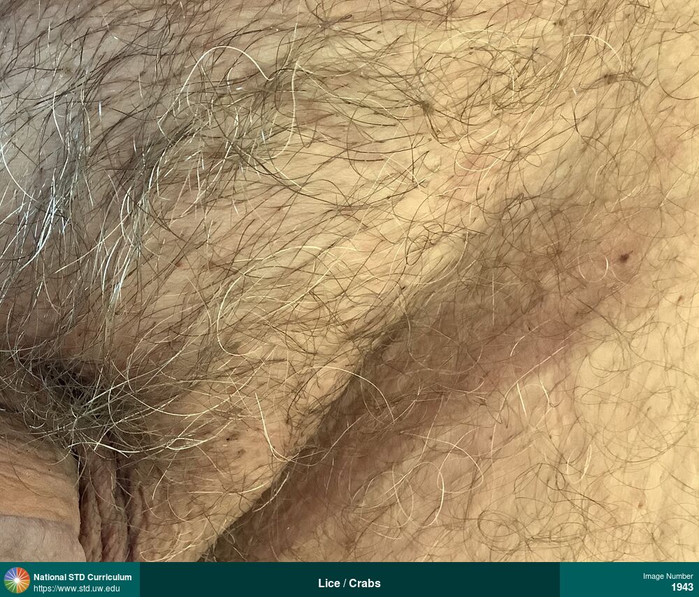
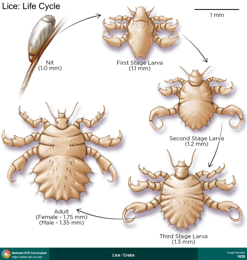
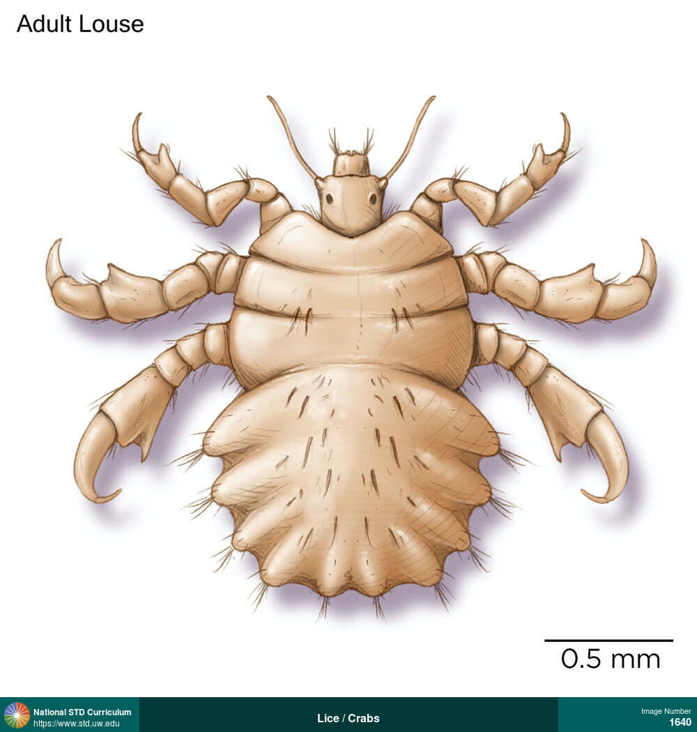
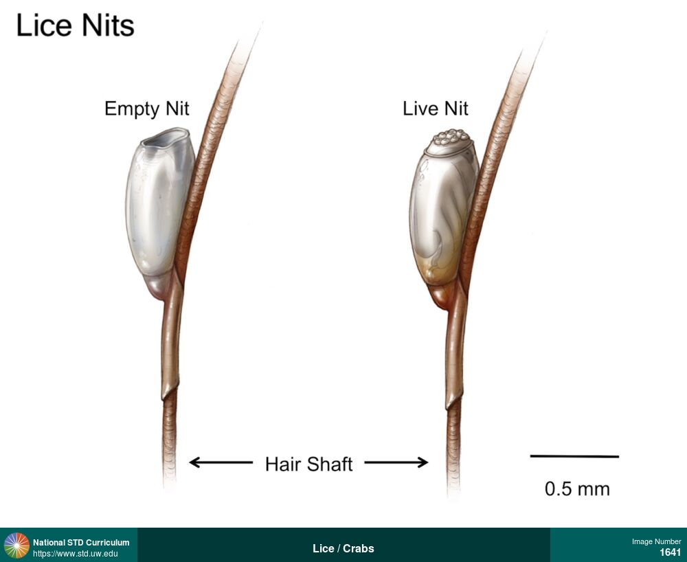
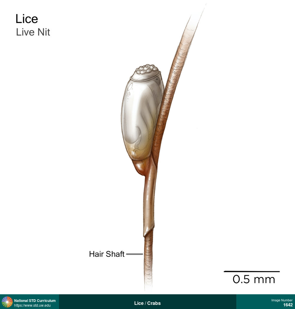
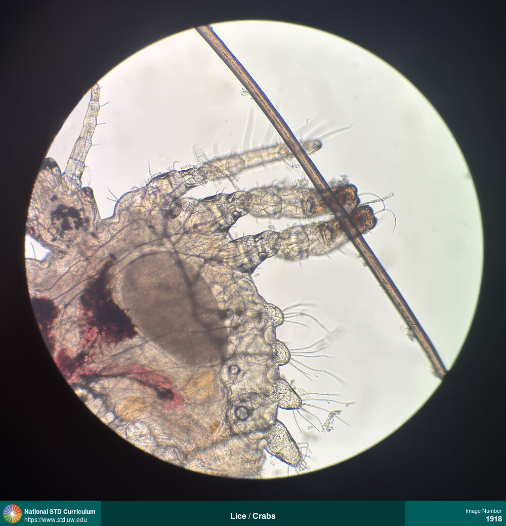
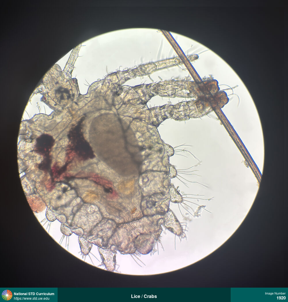
Lice / Crabs
This photograph shows the suprapubic region of a man with pubic lice (crab lice). The adult lice appear as tan-brown, slightly transparent oval-shaped objects and the nits appear as very small, oval-round dark dots attached to hair shafts.
Photo: Light skin tone, Suprapubic (Hypogastrium), Itch
Courtesy of Negusse Ocbamichael, PA
N/A Light skin tone, Suprapubic (Hypogastrium)
1943
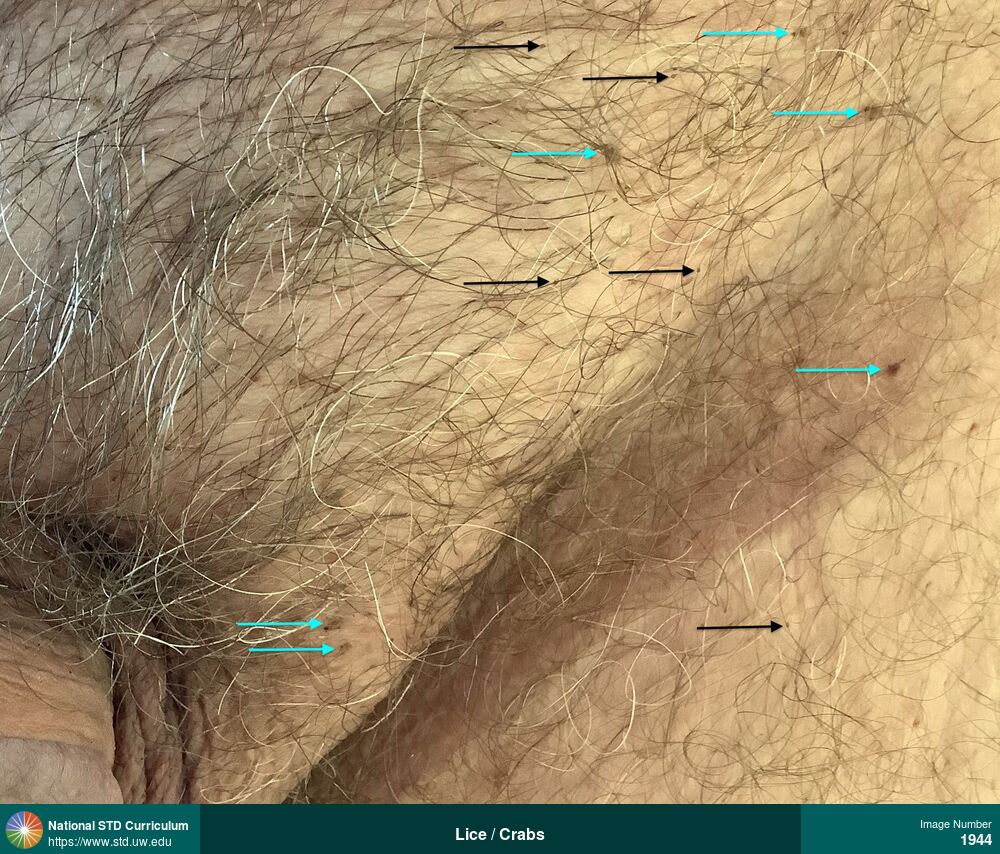
Lice / Crabs
This photograph shows the suprapubic region of a man with pubic lice (crab lice). The adult lice appear as tan-brown, slightly transparent oval-shaped objects (blue arrows) and the nits appear as very small, oval-round, dark dots attached to hair shafts (black arrows).
Photo: Light skin tone, Suprapubic (Hypogastrium), Itch
Courtesy of Negusse Ocbamichael, PA
N/A Light skin tone, Suprapubic (Hypogastrium)
1944

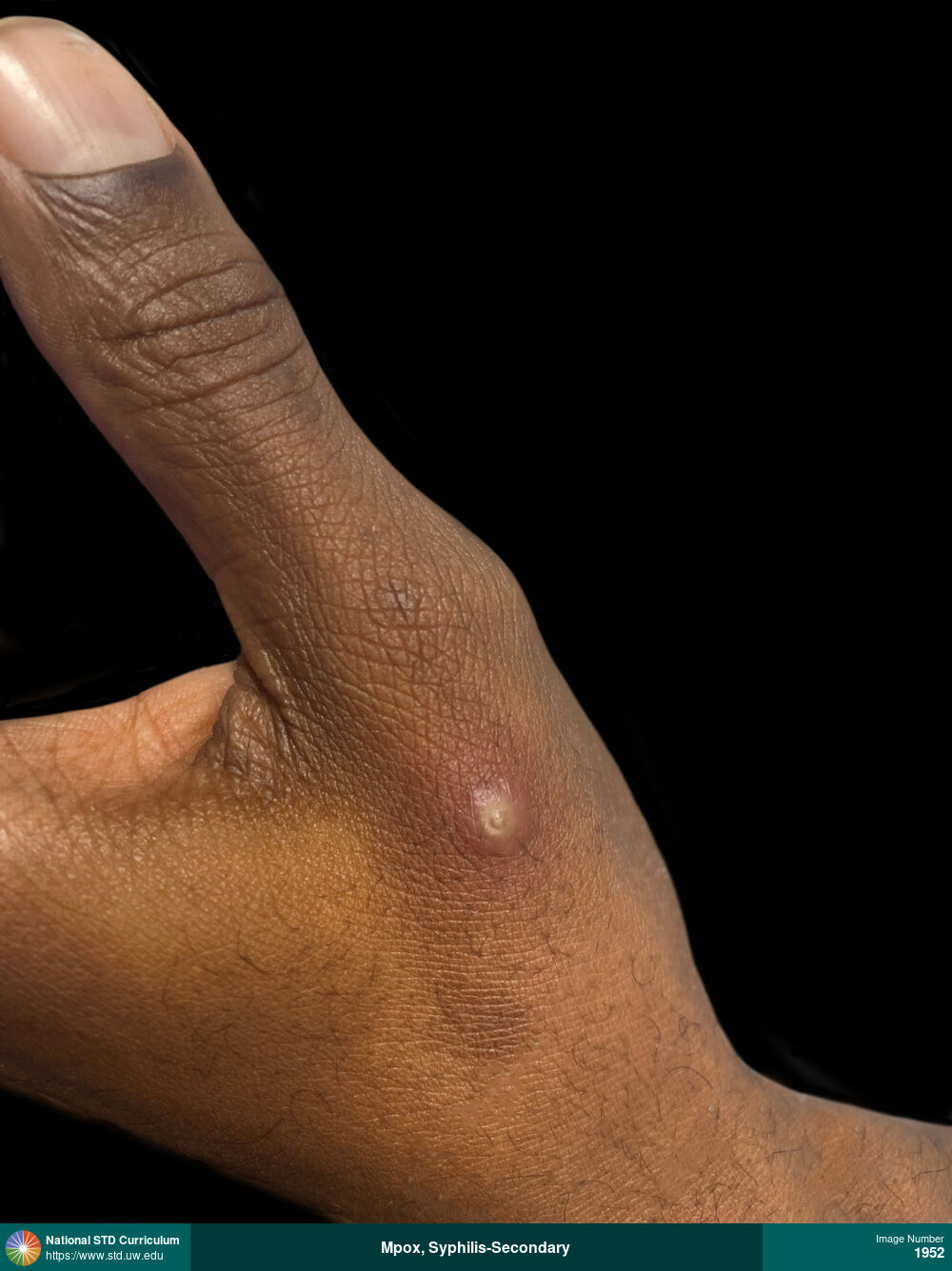
Mpox, Syphilis-Secondary
Pustular mpox lesion at base of left thumb.
Photo: Pustule / Pustules, Dark skin tone, Hand (Left)
Courtesy of Laura A. Quilter, MD, MPH
Pustule / Pustules Dark skin tone, Hand (Left)
1952
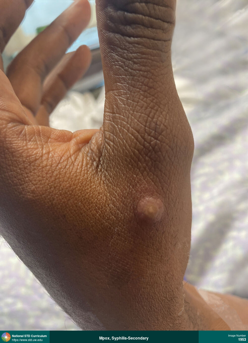
Mpox, Syphilis-Secondary
Pustlar mpox lesion on the base of the left thumb.
Photo: Pustule / Pustules, Rash, Dark skin tone, Hand (Left)
Courtesy of Laura A. Quilter, MD, MPH
Pustule / Pustules, Rash Dark skin tone, Hand (Left)
1953
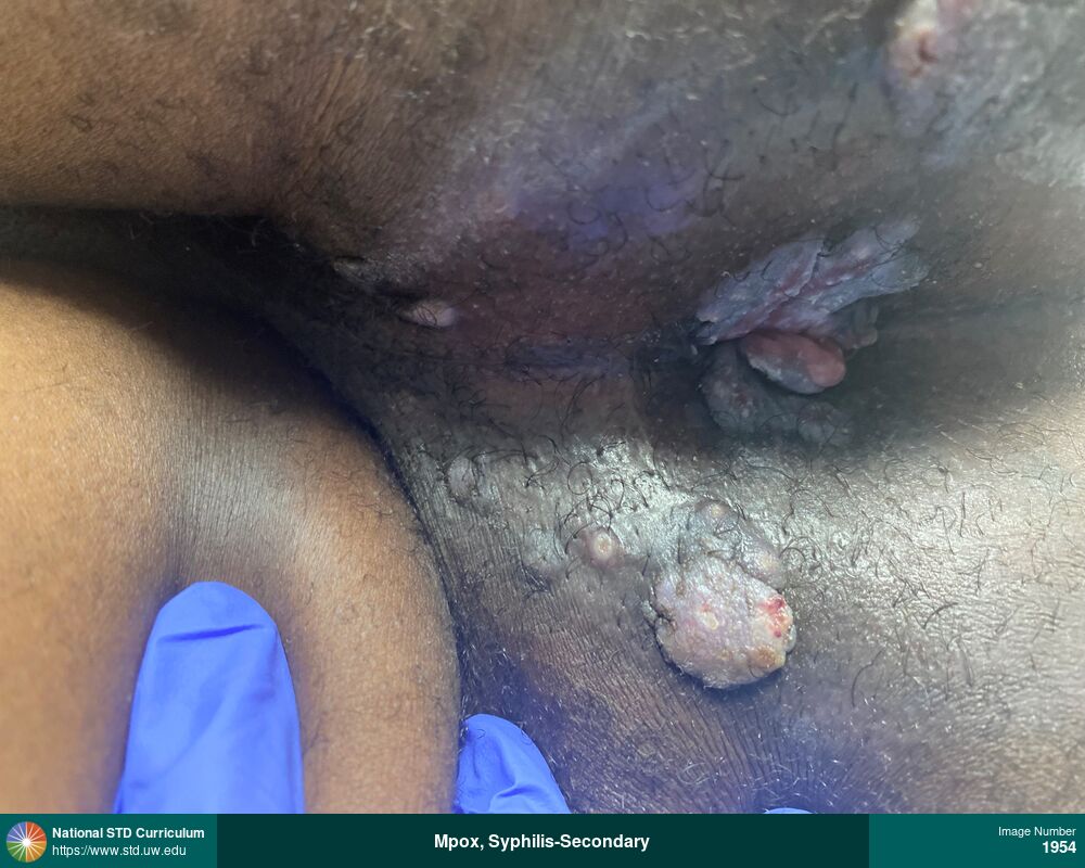
Mpox, Syphilis-Secondary
Perinal and gluteal cleft condylomata lata lesions caused by secondary syphilis. In addition, there is an umbilicated pustular mpox lesion.
Photo: Plaque, Pustule / Pustules, Verrucous, Anal / Perianal, Buttock, Dark skin tone, Rash
Courtesy of Laura A. Quilter, MD, MPH
Plaque, Pustule / Pustules, Verrucous Anal / Perianal, Buttock, Dark skin tone
1954
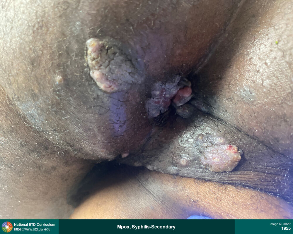
Mpox, Syphilis-Secondary
Perianal and gluteal cleft condylomata lata lesions. There are also several umbilicated pustular mpox lesions.
Photo: Plaque, Pustule / Pustules, Verrucous, Anal / Perianal, Buttock, Dark skin tone, Rash
Courtesy of Laura A. Quilter, MD, MPH
Plaque, Pustule / Pustules, Verrucous Anal / Perianal, Buttock, Dark skin tone
1955

Mpox, Syphilis-Secondary
Seconeary syphilis with multiple condylomata lata lesions in perianal and gluteal cleft region.
Photo: Plaque, Verrucous, Anal / Perianal, Buttock, Dark skin tone, Rash
Courtesy of Laura A. Quilter, MD, MPH
Plaque, Verrucous Anal / Perianal, Buttock, Dark skin tone
1956
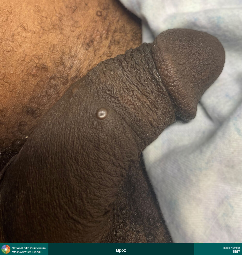
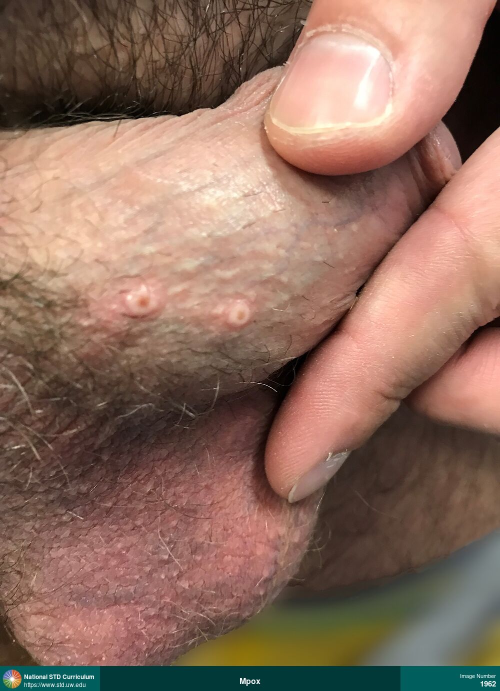
Mpox, Syphilis-Secondary
Pustular umbilicated papular mpox lesion on shaft of penis.
Photo: Papule / Papules, Dark skin tone, Penis
Courtesy of Laura A. Quilter, MD, MPH
Papule / Papules Dark skin tone, Penis
1957
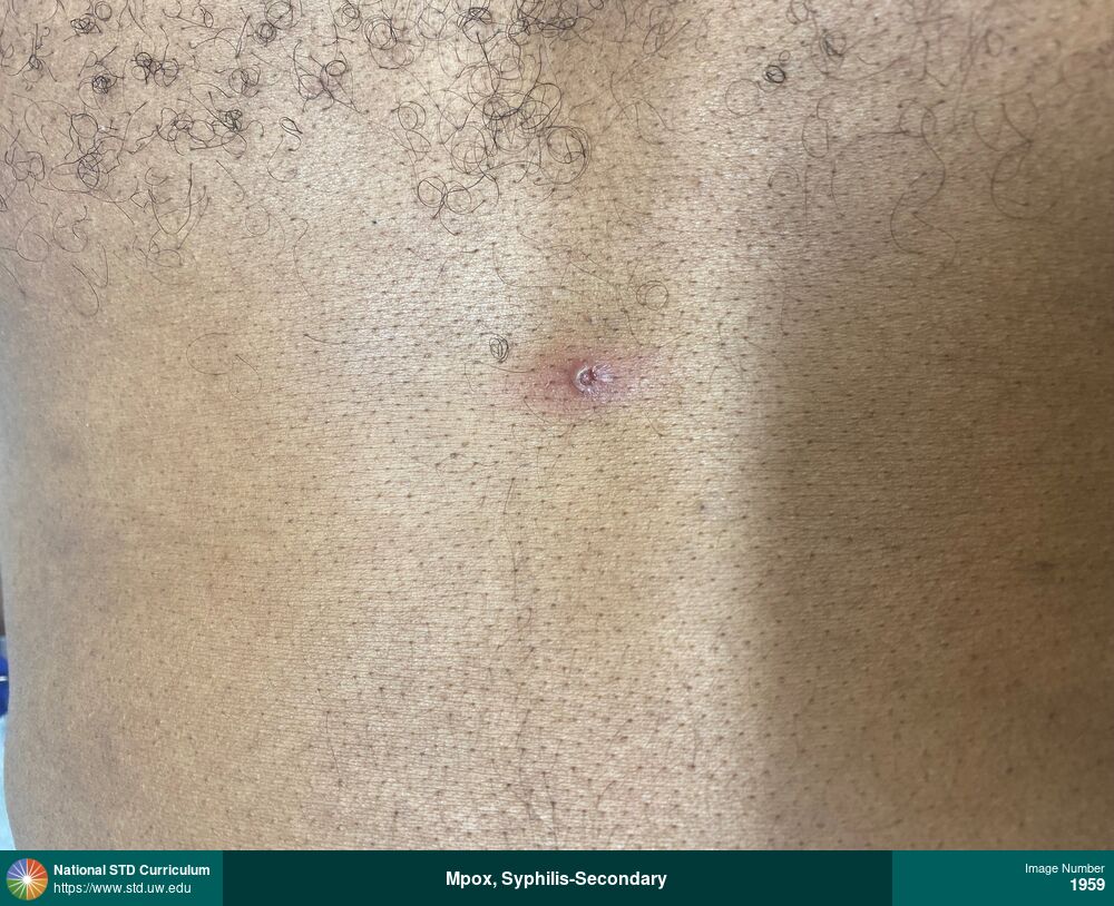
Mpox, Syphilis-Secondary
Umbilicated pustular mpox lesion, with surrounding erythema, on chest
Photo: Erythema, Pustule / Pustules, Chest, Dark skin tone
Courtesy of Laura A. Quilter, MD, MPH
Erythema, Pustule / Pustules Chest, Dark skin tone
1959
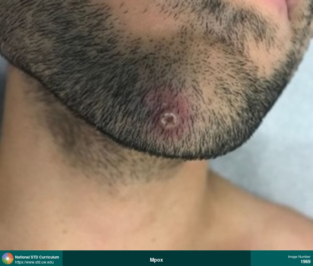
Mpox
Umbilicated pustular mpox lesions at base of penile shaft.
Photo: Pustule / Pustules, Light skin tone, Penis, Rash
Courtesy of Bryce Furness, MD, MPH
Pustule / Pustules Light skin tone, Penis
1962

Mpox
Multiple vesicular-pustular mpox lesions on shaft of penis.
Photo: Pustule / Pustules, Vesicle / Vesicles, Light skin tone, Penis, Rash
Courtesy of Bryce Furness, MD, MPH
Pustule / Pustules, Vesicle / Vesicles Light skin tone, Penis
1963
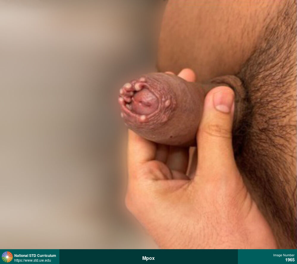
Mpox
Multiple pustular mpox lesions on the foreskin of the penis.
Photo: Pustule / Pustules, Light skin tone, Penis, Painful
Courtesy of Bryce Furness, MD, MPH
Pustule / Pustules Light skin tone, Penis
1965
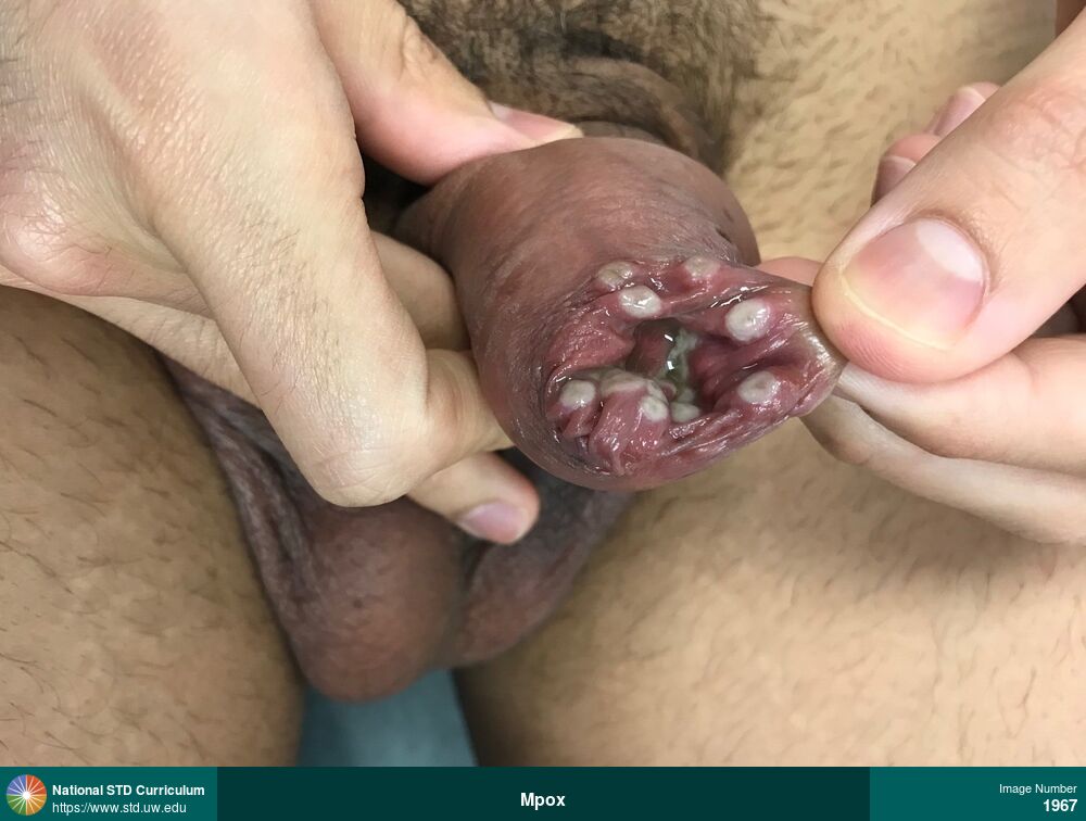
Mpox
Extensive pustular mpox lesions on foreskin of penis associated with edema of foreskin
Photo: Pustule / Pustules, Rash, Light skin tone, Penis, Painful, Rash
Courtesy of Bryce Furness, MD, MPH
Pustule / Pustules, Rash Light skin tone, Penis
1967
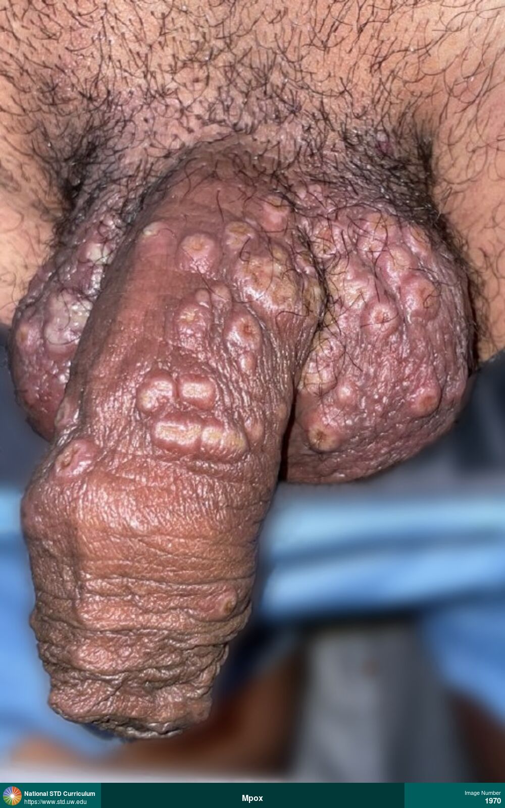
Mpox
Multiple pustular and umbilicated mpox lesions located bilaterally on the scrotum and along the penile shaft and foreskin.
Photo: Pustule / Pustules, Umbilicated, Vesicle / Vesicles, Dark skin tone, Penis, Scrotum
Courtesy of Mireya A. Wessolossky, MD
Pustule / Pustules, Umbilicated, Vesicle / Vesicles Dark skin tone, Penis, Scrotum
1970
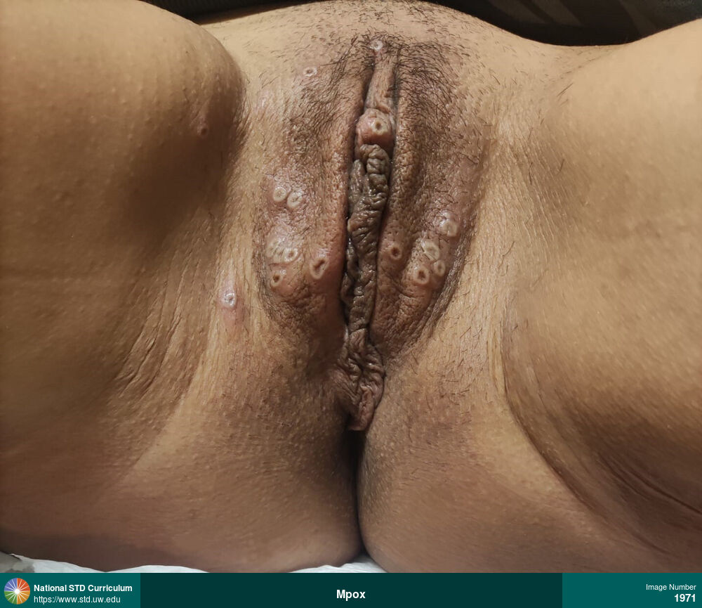
Mpox
Photo: Pustule / Pustules, Rash, Verrucous, Dark skin tone, Labia (majora/minora), Vulva, Non-Painful, Rash
Courtesy of Joana Portela Dias, MD
Pustule / Pustules, Rash, Verrucous Dark skin tone, Labia (majora/minora), Vulva
1971
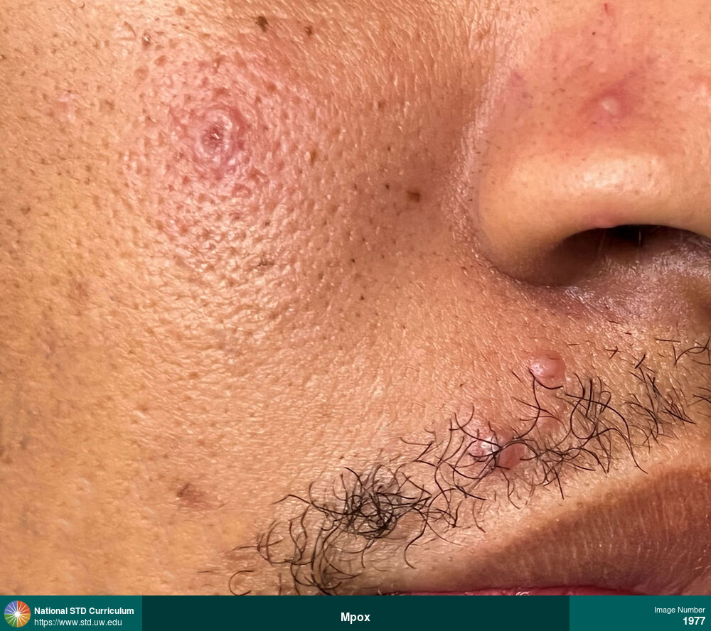
Mpox
Mpox lesion in early stages on face, with papular and vesicular lesions.
Photo: Papule / Papules, Vesicle / Vesicles, Dark skin tone, Face, Painful
Courtesy of Laura A. Quilter, MD, MPH
Papule / Papules, Vesicle / Vesicles Dark skin tone, Face
1977
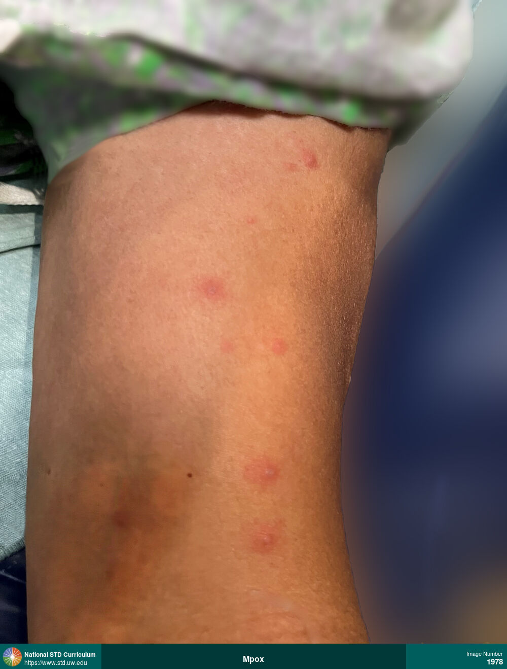
Mpox
Mpox lesion in early stages, including macular lesions, papular lesions, and small vesicular lesions.
Photo: Macule / Macules, Papule / Papules, Vesicle / Vesicles, Dark skin tone
Courtesy of Laura A. Quilter, MD, MPH
Macule / Macules, Papule / Papules, Vesicle / Vesicles Dark skin tone
1978
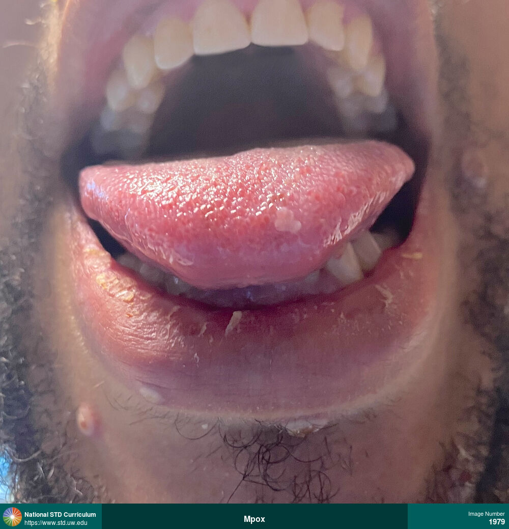
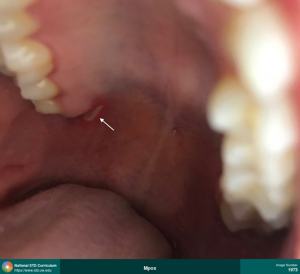
Mpox
Mpox lesion on the tip of the tongue and other lesions on the lower lip and face.
Photo: Plaque, Pustule / Pustules, Vesicle / Vesicles, Dark skin tone, Lips, Tongue, Painful
Courtesy of Laura A. Quilter, MD, MPH
Plaque, Pustule / Pustules, Vesicle / Vesicles Dark skin tone, Lips, Tongue
1979
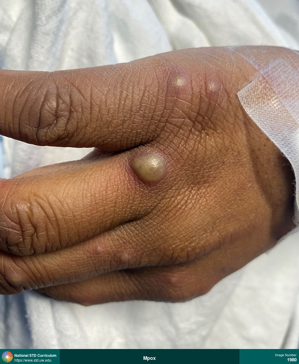
Mpox
A large, bullous mpox lesion at the base of a finger, with several smaller papular and vesicular mpox lesions on the hand.
Photo: Bulla / Bullae, Pustule / Pustules, Vesicle / Vesicles, Dark skin tone, Hand (Left), Painful
Courtesy of Laura A. Quilter, MD, MPH
Bulla / Bullae, Pustule / Pustules, Vesicle / Vesicles Dark skin tone, Hand (Left)
1980
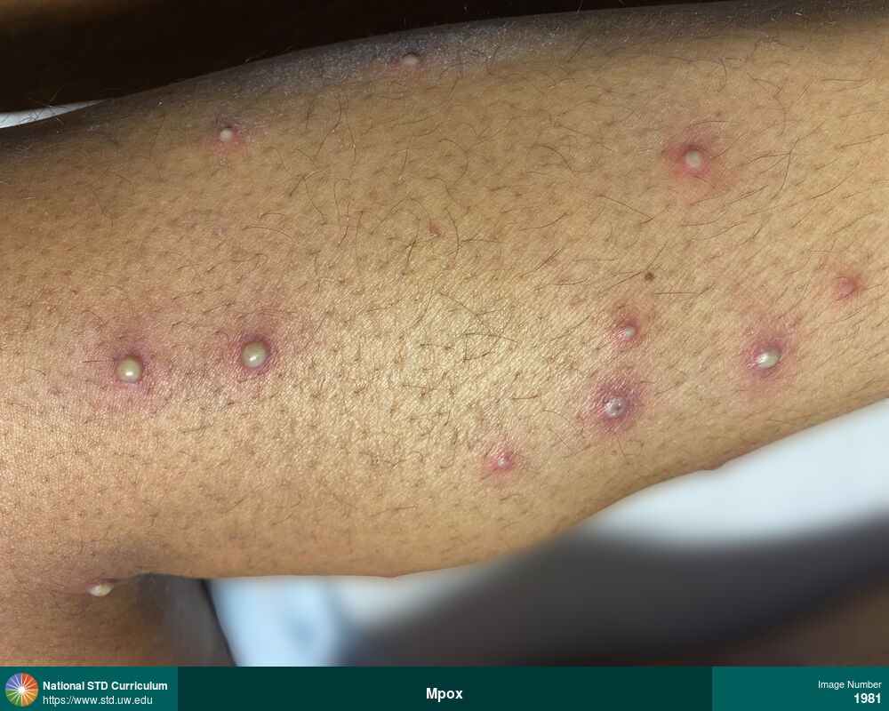
Mpox
Close-up view of pustular mpox lesions on lower posterior leg region.
Photo: Pustule / Pustules, Calf, Dark skin tone
Courtesy of Laura A. Quilter, MD, MPH
Pustule / Pustules Calf, Dark skin tone
1981
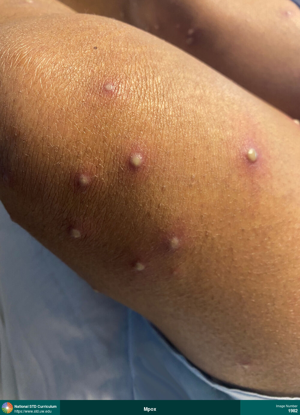
Mpox
Close-up view of pustular mpox lesions on the upper left thigh region.
Photo: Pustule / Pustules, Dark skin tone, Thigh
Courtesy of Laura A. Quilter, MD, MPH
Pustule / Pustules Dark skin tone, Thigh
1982
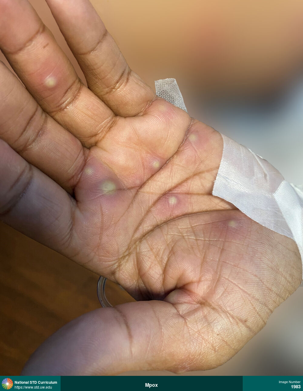
Mpox
Mpox vescicular and pustular lesions on the left palm.
Photo: Pustule / Pustules, Vesicle / Vesicles, Dark skin tone, Hand (Left), Painful
Courtesy of Laura A. Quilter, MD, MPH
Pustule / Pustules, Vesicle / Vesicles Dark skin tone, Hand (Left)
1983
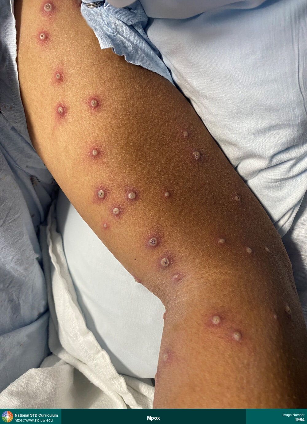
Mpox
Extensive pustular mpox lesions on the left upper arm.
Photo: Pustule / Pustules, Arm (Left), Dark skin tone, Painful
Courtesy of Laura A. Quilter, MD, MPH
Pustule / Pustules Arm (Left), Dark skin tone
1984

Mpox
Mpox lesions on the face and periorbital region, include eyelid lesion with secondary eyelid edema.
Photo: Pustule / Pustules, Vesicle / Vesicles, Dark skin tone, Eye, Face, Painful
Courtesy of Laura A. Quilter, MD, MPH
Pustule / Pustules, Vesicle / Vesicles Dark skin tone, Eye, Face
1985
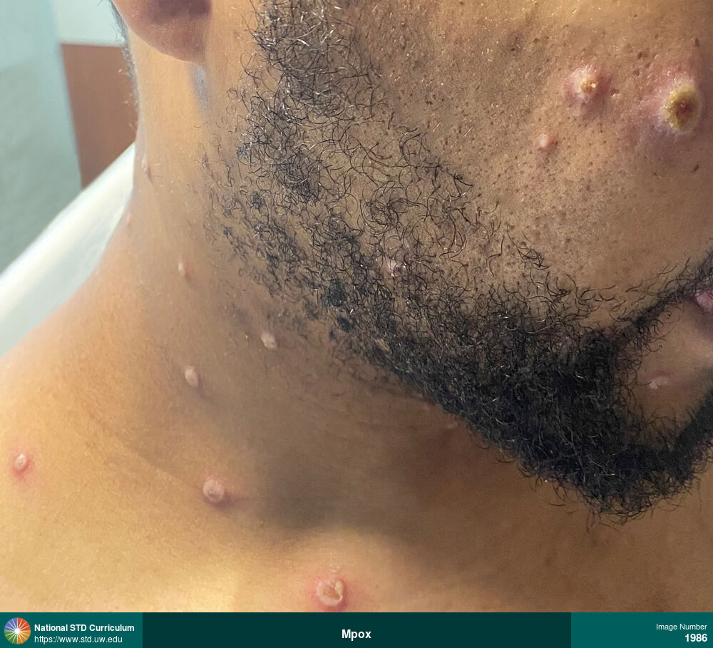
Mpox
Mpox lesions of different stages and sizes on upper chest, neck, and face.
Photo: Pustule / Pustules, Vesicle / Vesicles, Dark skin tone, Face, Neck, Trunk/Torso, Painful
Courtesy of Laura A. Quilter, MD, MPH
Pustule / Pustules, Vesicle / Vesicles Dark skin tone, Face, Neck, Trunk/Torso
1986
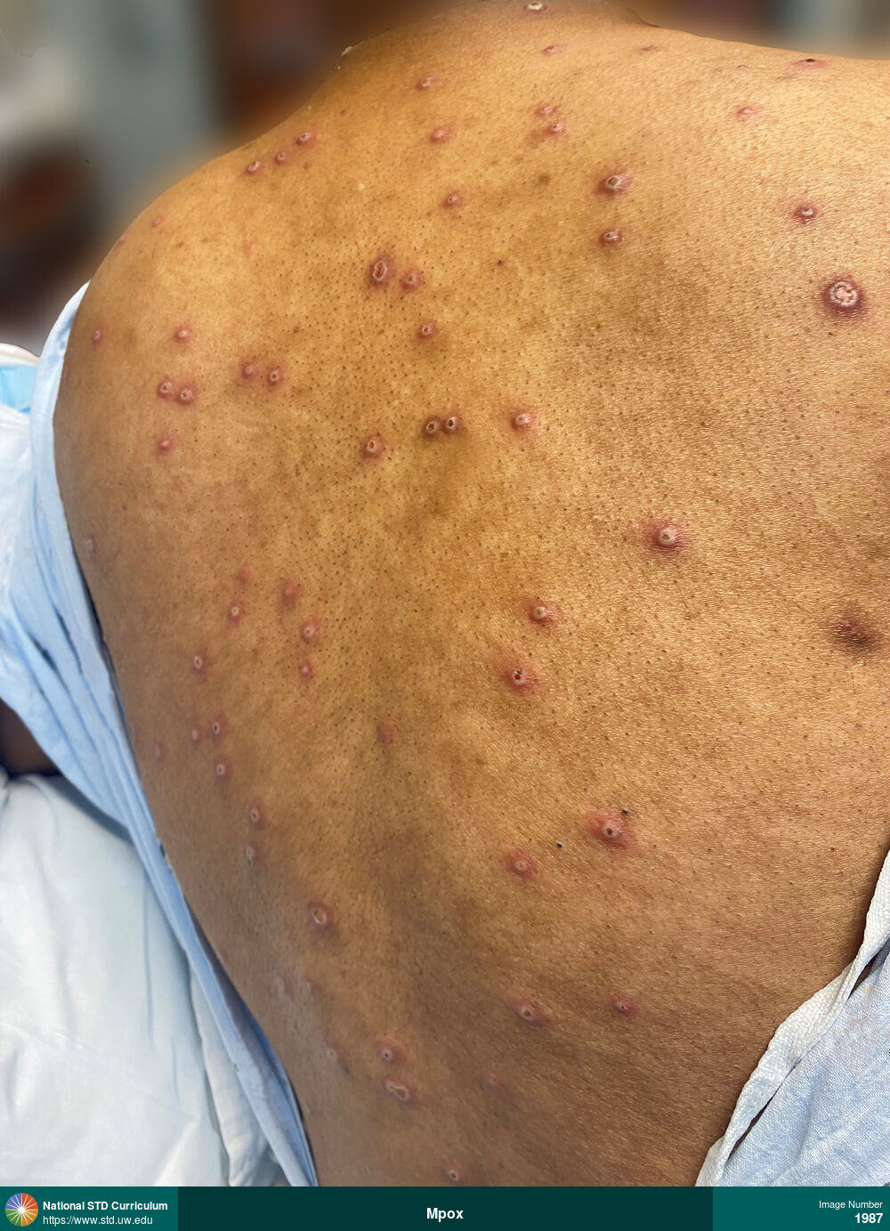
Mpox
Extensive, large pustular mpox lesions on back.
Photo: Pustule / Pustules, Back, Dark skin tone
Courtesy of Laura A. Quilter, MD, MPH
Pustule / Pustules Back, Dark skin tone
1987
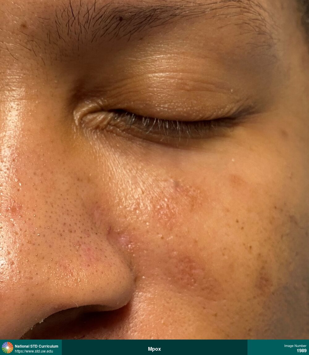
Mpox
Healed mpox lesions on face.
Photo: Healed lesions, Dark skin tone, Face, Non-Painful
Courtesy of Laura A. Quilter, MD, MPH
Healed lesions Dark skin tone, Face
1989
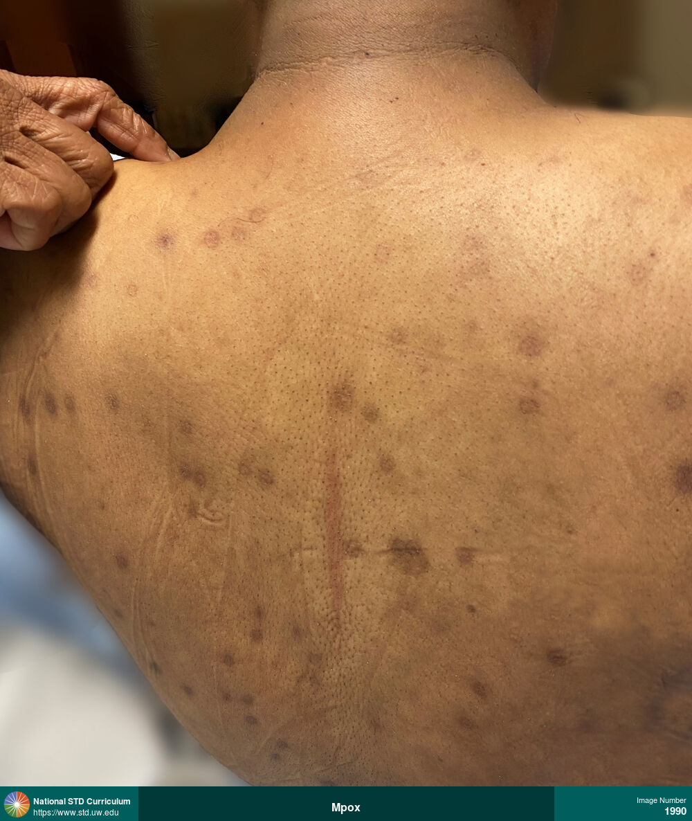
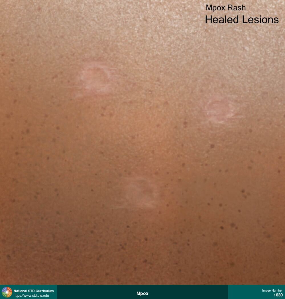
Mpox
Healed mpox lesions on back.
Photo: Healed lesions, Back, Dark skin tone
Courtesy of Laura A. Quilter, MD, MPH
Healed lesions Back, Dark skin tone
1990
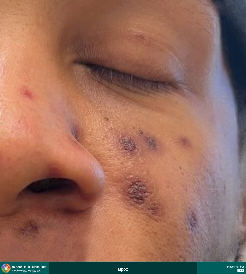
Mpox
Extensive pustular mpox lesions on the face, with variable stages and size of lesions.
Photo: Pustule / Pustules, Vesicle / Vesicles, Dark skin tone, Face, Painful
Courtesy of Laura A. Quilter, MD, MPH
Pustule / Pustules, Vesicle / Vesicles Dark skin tone, Face
1992
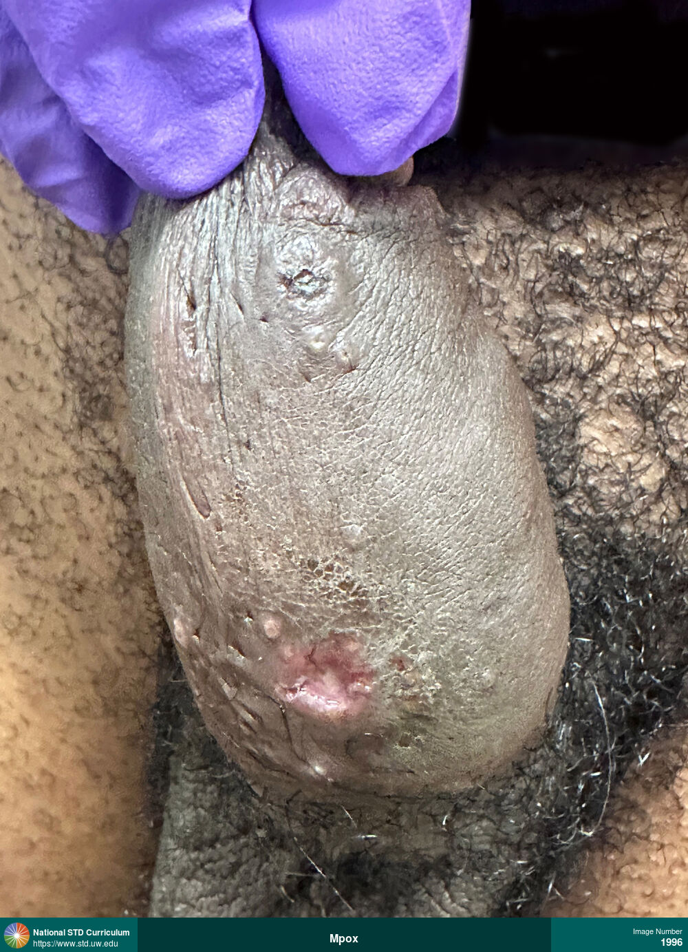
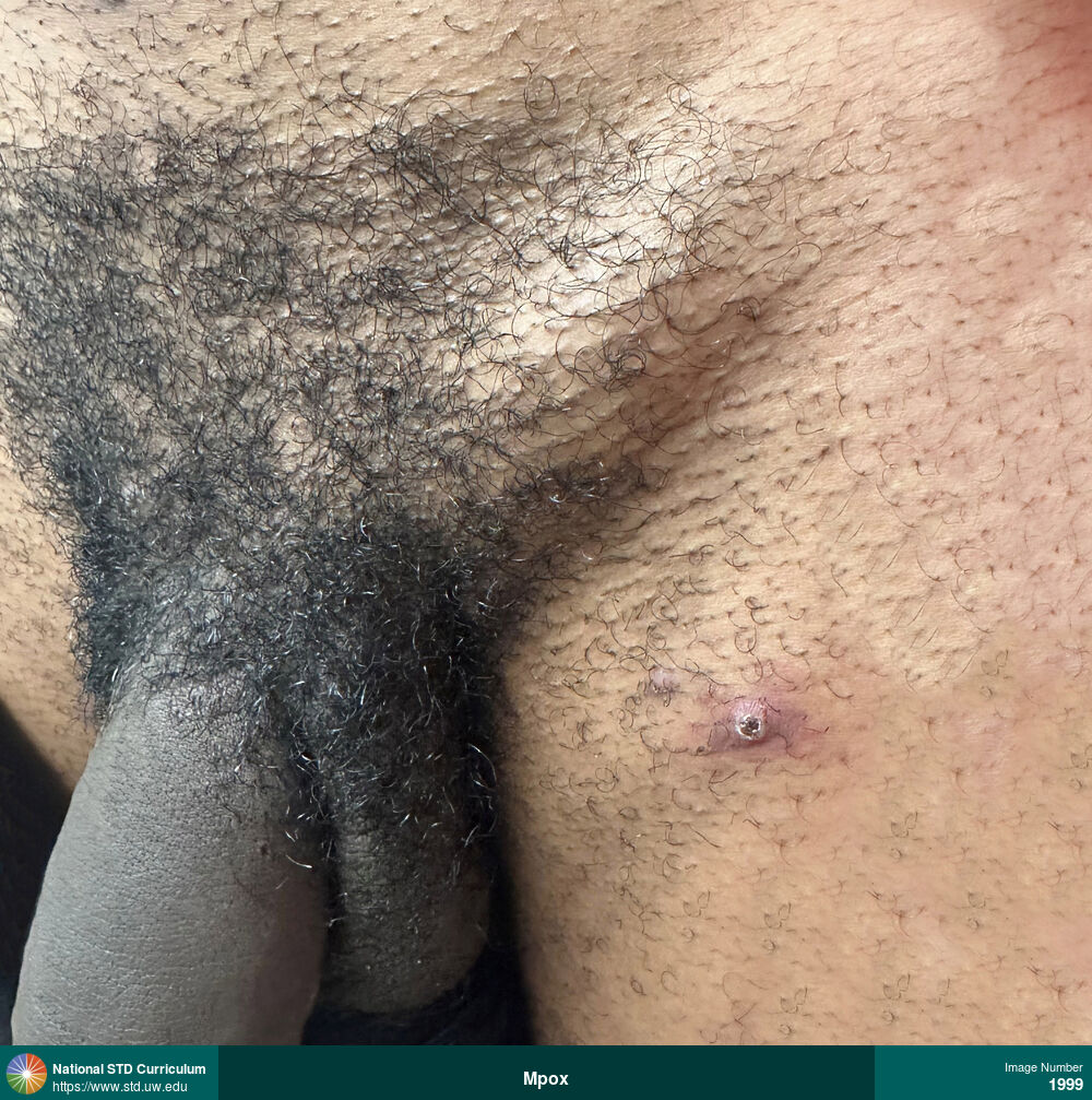
Mpox
Large, ulcerated, painful mpox lesion at the base of the penis.
Photo: Ulcer / Ulcers, Dark skin tone, Penis
Courtesy of Laura A. Quilter, MD, MPH
Ulcer / Ulcers Dark skin tone, Penis
1996
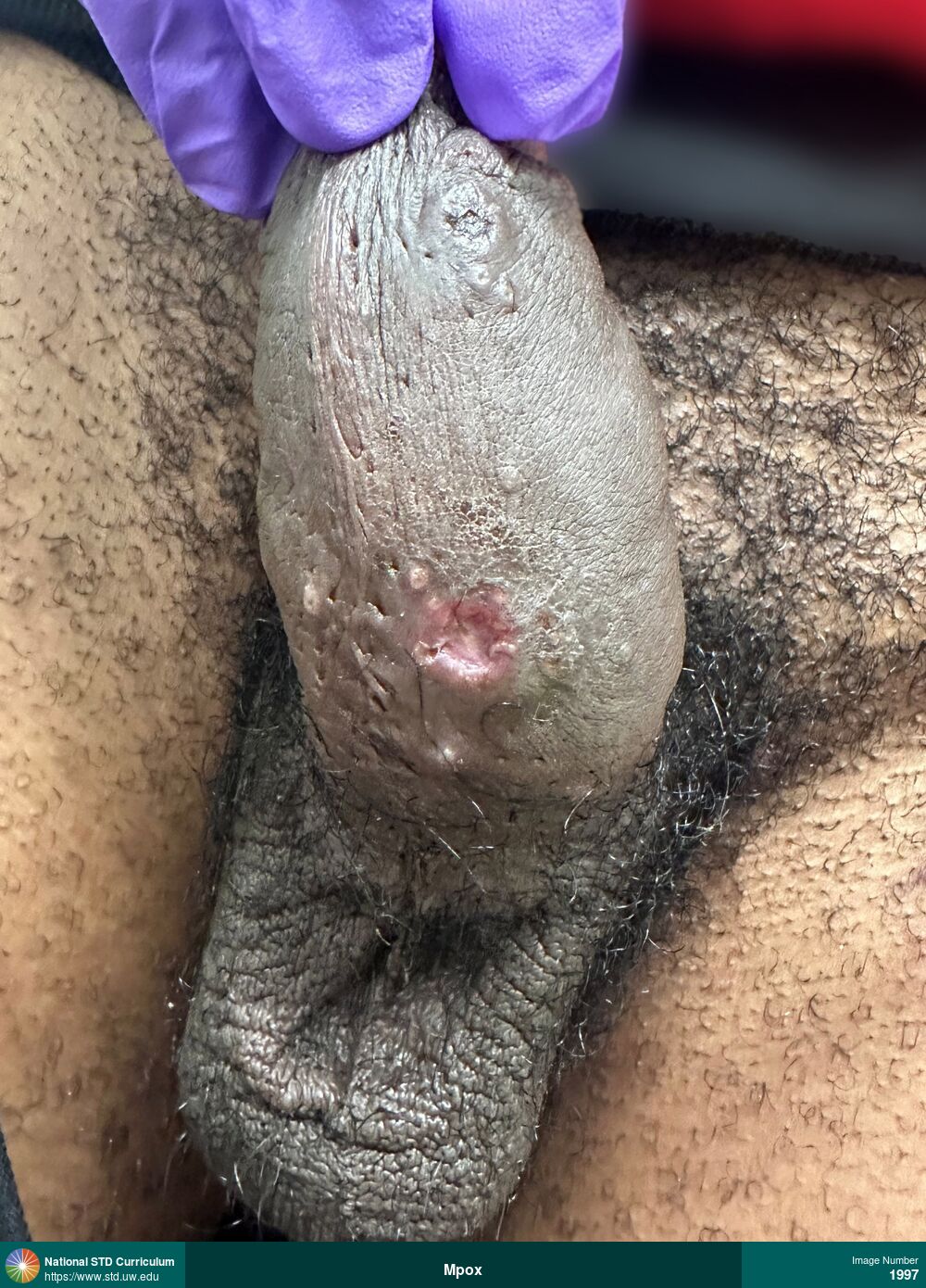
Mpox
Large, ulcerated, painful mpox lesion at the base of the penis.
Photo: Ulcer / Ulcers, Dark skin tone, Penis, Painful
Courtesy of Laura A. Quilter, MD, MPH
Ulcer / Ulcers Dark skin tone, Penis
1997
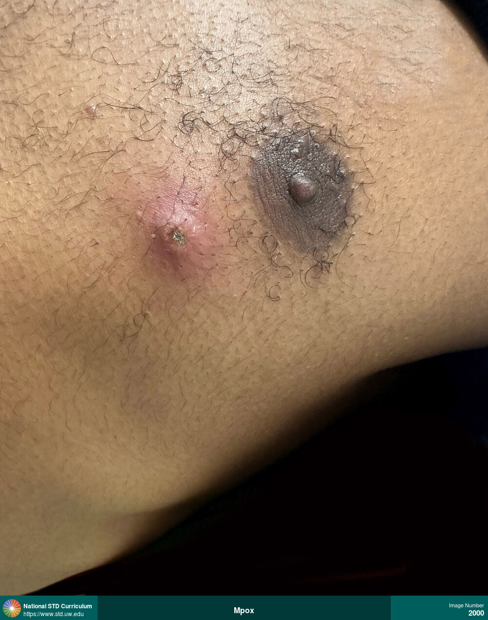
Mpox
Erythematous, firm, umbilicated solitary mpox lesion on left chest inferior to left nipple. The lesion was pruritic, tender, and painful.
Photo: Ulcer / Ulcers, Umbilicated, Vesicle / Vesicles, Chest, Dark skin tone, Itch, Painful
Courtesy of Laura A. Quilter, MD, MPH
Ulcer / Ulcers, Umbilicated, Vesicle / Vesicles Chest, Dark skin tone
2000
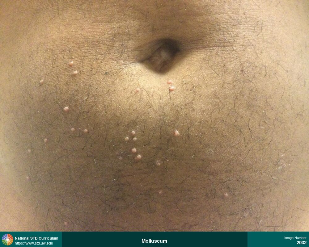
Molluscum
Multiple small, umbilicated papules on the lower abdominal region caused by molluscum contagiosum.
Photo: Papule / Papules, Umbilicated, Abdomen, Suprapubic (Hypogastrium), Non-Itchy, Non-Painful
Courtesy of Ben Garfinkel, PA-C
Papule / Papules, Umbilicated Abdomen, Suprapubic (Hypogastrium)
2032

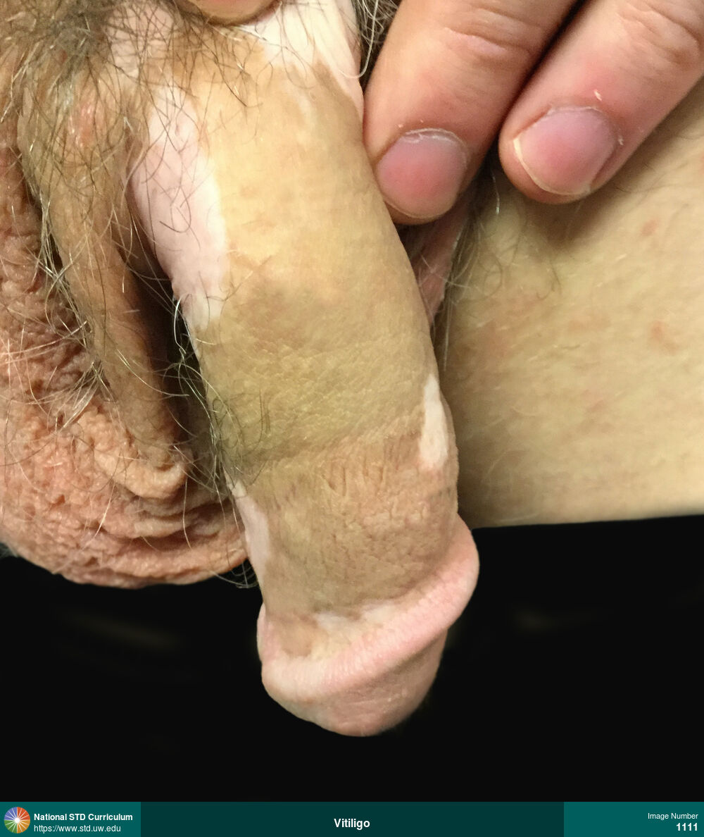
Vitiligo
Hypopigmented macular patches involving the dorsal region of the glans penis.
Photo: Hypopigmentation, Macule / Macules, Dark skin tone, Penis, Non-Itchy, Non-Painful
Courtesy of Ben Garfinkel, PA-C
Hypopigmentation, Macule / Macules Dark skin tone, Penis
2034
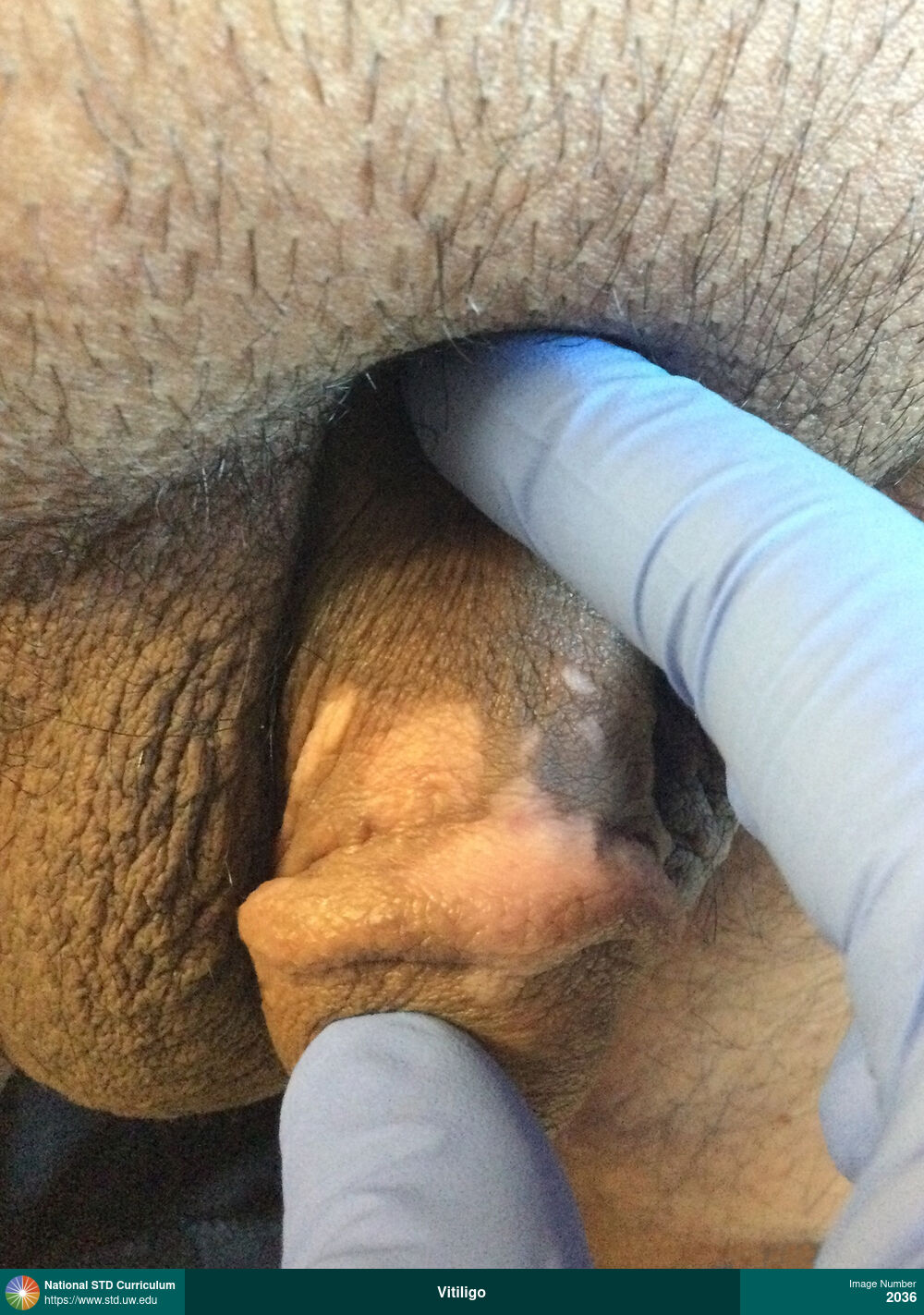
Vitiligo
Hypopigmented macular patches involving the dorsal shaft of the penis and a small region of the glans.
Photo: Hypopigmentation, Macule / Macules, Dark skin tone, Penis, Non-Itchy, Non-Painful
Courtesy of Ben Garfinkel, PA-C
Hypopigmentation, Macule / Macules Dark skin tone, Penis
2036
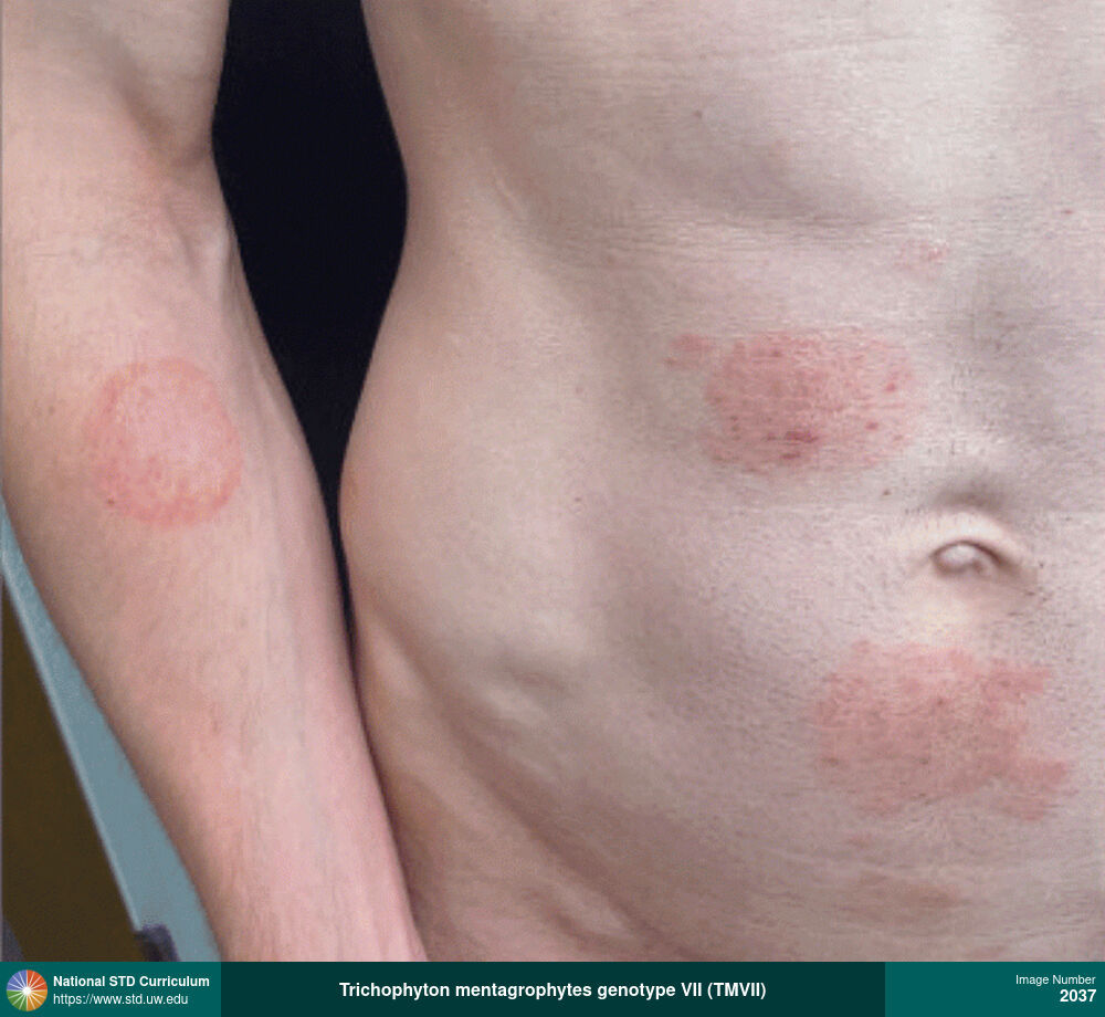
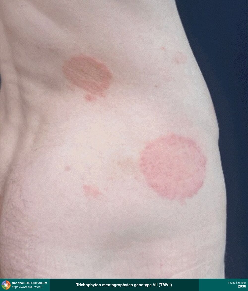
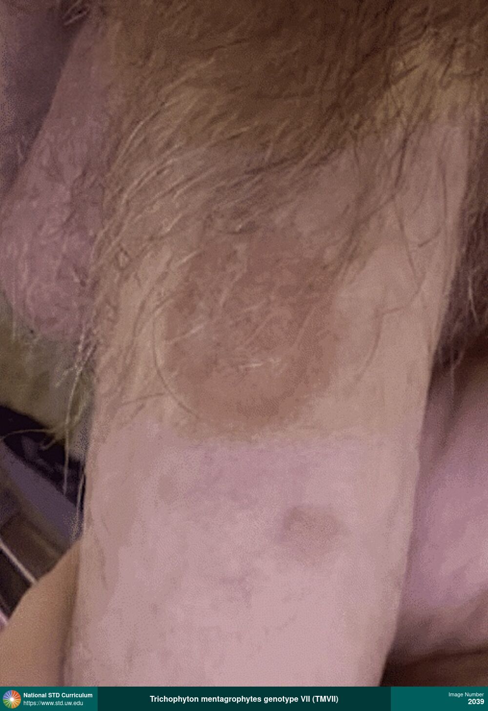
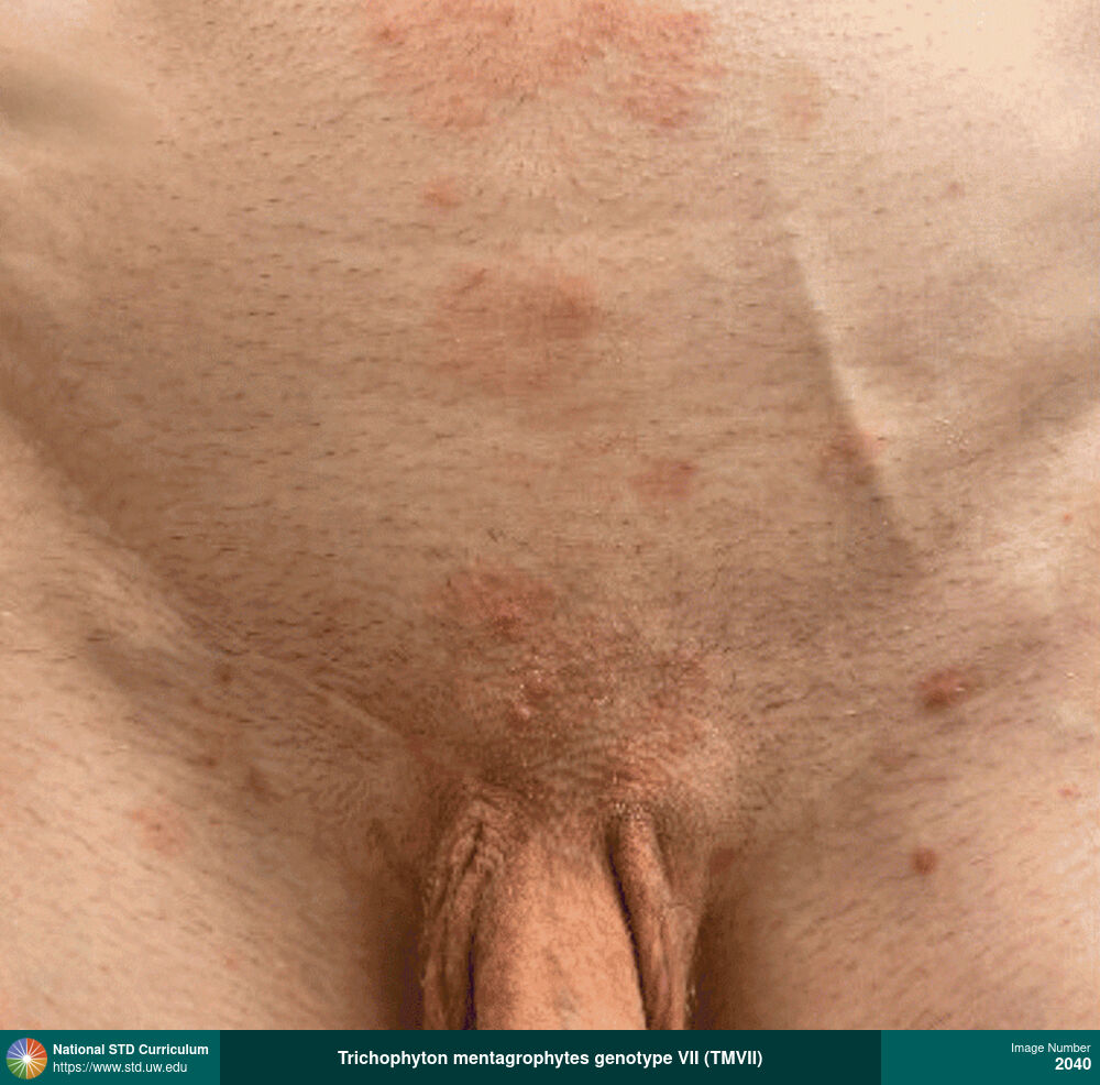
Trichophyton mentagrophytes genotype VII (TMVII)
Multiple, annular, scaly plaques on the right arm and lower abdominal region in a man who reported sex with other men. A skin scraping placed in fungal culture grew organisms identified as Trichophyton mentagrophytes genotype VII based on genetic sequencing. The rash did not respond to initial treatment with topical therapy (clotrimazole and then terbinafine) but subsequently responded to oral terbinafine. The authors conclude the fungal infection was likely sexually transmitted.
Source: Zucker J, Caplan AS, Gunaratne SH, et al. Notes from the Field: Trichophyton mentagrophytes Genotype VII - New York City, April-July 2024. MMWR Morb Mortal Wkly Rep. 2024;73:985-8.
Source: Zucker J, Caplan AS, Gunaratne SH, et al. Notes from the Field: Trichophyton mentagrophytes Genotype VII - New York City, April-July 2024. MMWR Morb Mortal Wkly Rep. 2024;73:985-8.
Photo: Annular, Erythema, Plaque, Rash, Scale, Abdomen, Arm (Right), Light skin tone, Suprapubic (Hypogastrium), Rash
Courtesy of Avrom S. Caplan, MD
Annular, Erythema, Plaque, Rash, Scale Abdomen, Arm (Right), Light skin tone, Suprapubic (Hypogastrium)
2037
Trichophyton mentagrophytes genotype VII (TMVII)
Multiple, erythematous, scaly plaques on the lower abdominal and genital area, with scattered erythematous papulonodules (likely indicating follicular involvement of dermatophytosis) in a man who reported sex with other men. Organisms that grew in a fungal culture of a skin scraping were identified as Trichophyton mentagrophytes genotype VII based on genetic sequencing. The authors conclude the fungal infection was likely sexually transmitted.
Source: Zucker J, Caplan AS, Gunaratne SH, et al. Notes from the Field: Trichophyton mentagrophytes Genotype VII - New York City, April-July 2024. MMWR Morb Mortal Wkly Rep. 2024;73:985-8.
Source: Zucker J, Caplan AS, Gunaratne SH, et al. Notes from the Field: Trichophyton mentagrophytes Genotype VII - New York City, April-July 2024. MMWR Morb Mortal Wkly Rep. 2024;73:985-8.
Photo: Annular, Erythema, Plaque, Rash, Scale, Abdomen, Light skin tone, Suprapubic (Hypogastrium), Rash
Courtesy of Avrom S. Caplan, MD
Annular, Erythema, Plaque, Rash, Scale Abdomen, Light skin tone, Suprapubic (Hypogastrium)
2040
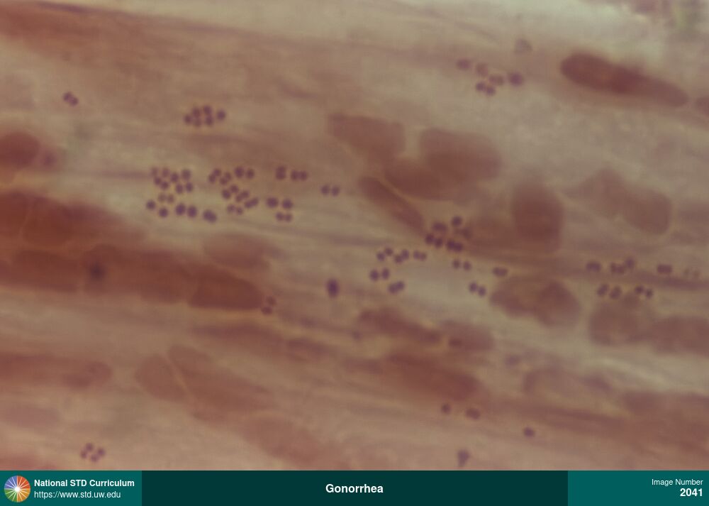
Gonorrhea
Gram stain of a rectal swab specimen from a patient with rectal gonorrhea showing multiple diplococci.
Photo: Discharge / Anal, Anal / Intra-anal, Microscope-Gram Stain, Rectal, Discharge, Painful
Courtesy of Ben Garfinkel, PA-C
Discharge / Anal Anal / Intra-anal, Microscope-Gram Stain, Rectal
2041
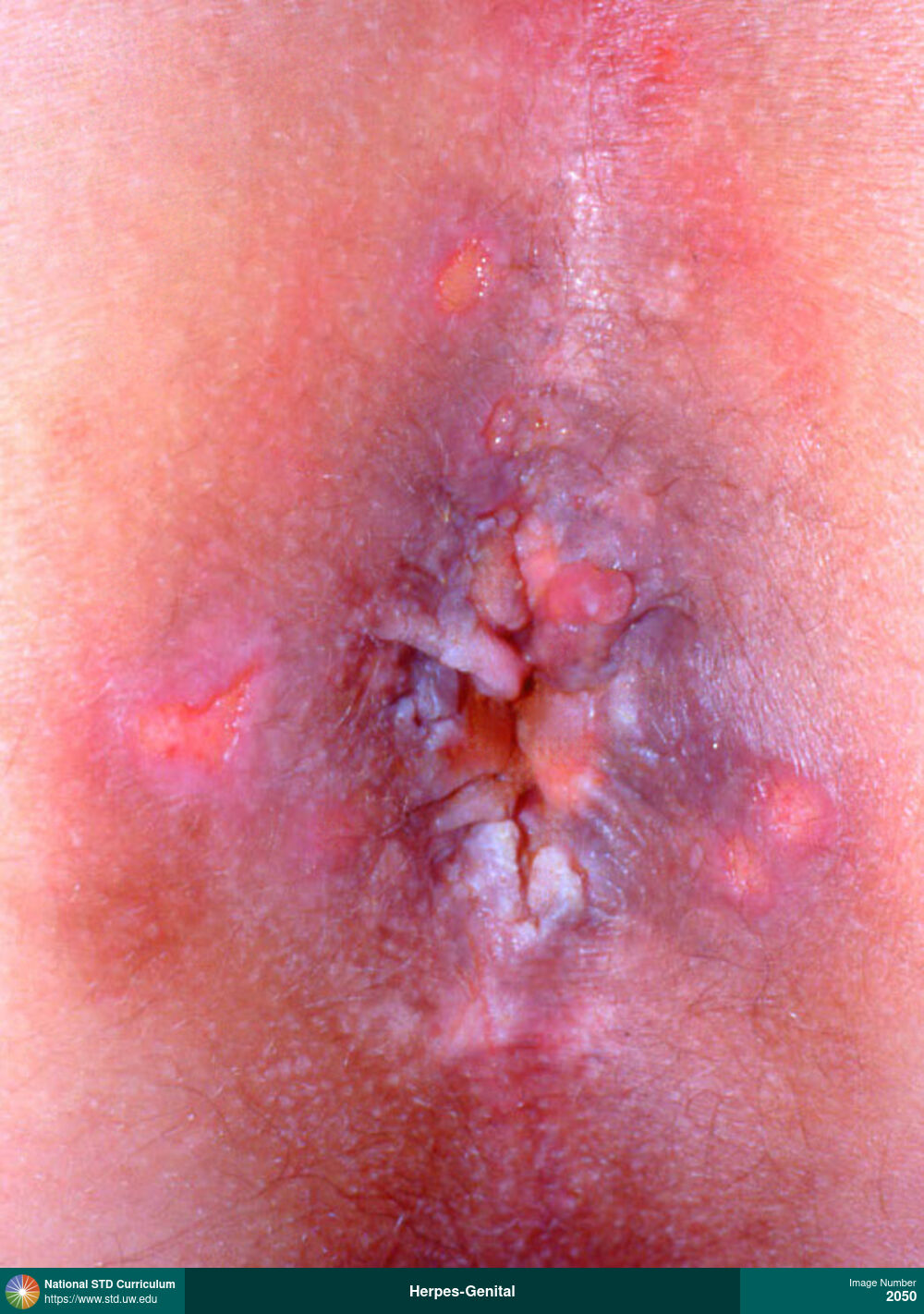
Herpes-Genital
Multiple, discrete, painful perianal ulcers in a man with HIV.
Photo: Ulcer / Ulcers, Anal / Perianal, Light skin tone, Painful
Courtesy of David H. Spach, MD
Ulcer / Ulcers Anal / Perianal, Light skin tone
2050
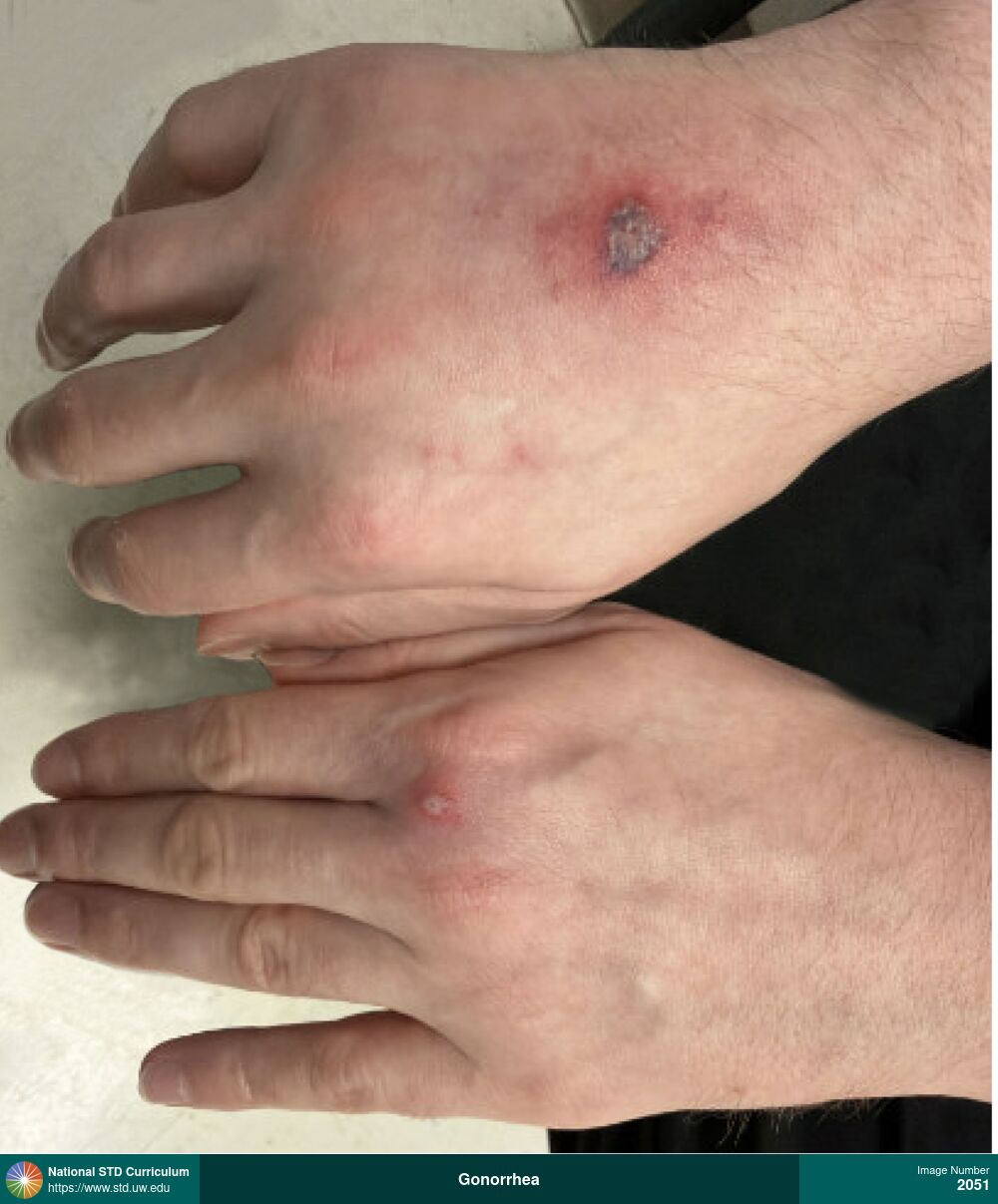
Gonorrhea
Disseminated gonococcal infection with arthritis
Photo: Pustule / Pustules, Scab, Ulcer / Ulcers, Arm (Left), Arm (Right), Light skin tone
Pustule / Pustules, Scab, Ulcer / Ulcers Arm (Left), Arm (Right), Light skin tone
2051
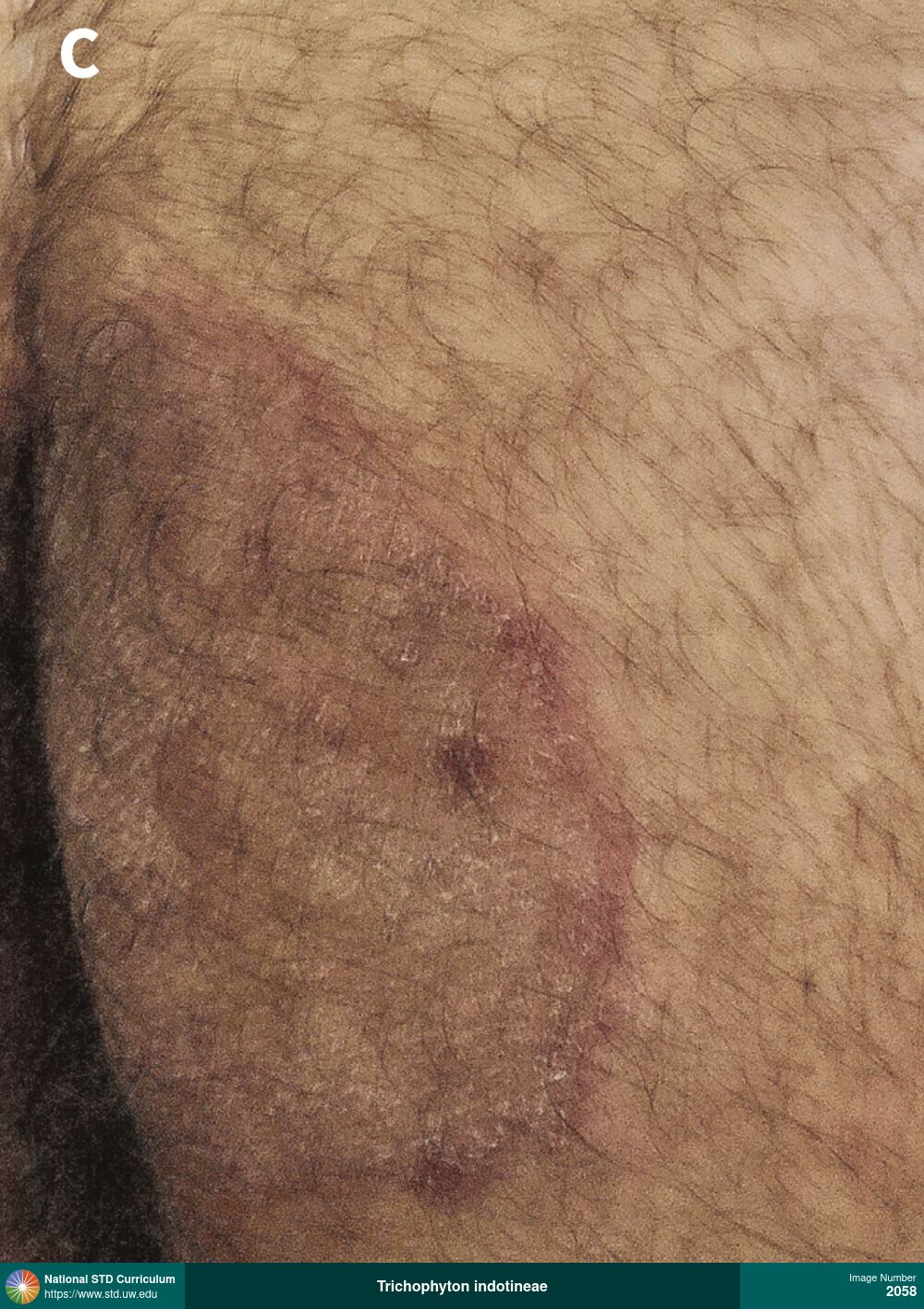
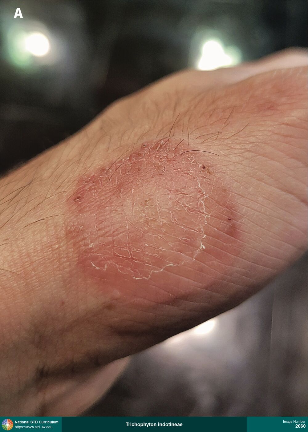
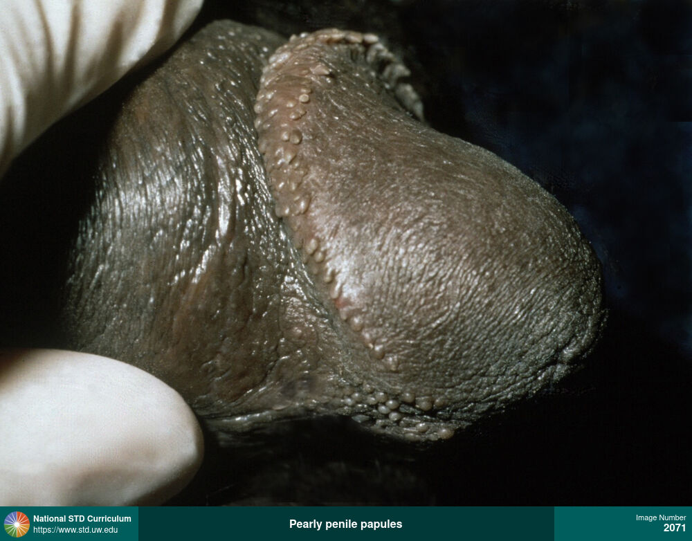
Pearly penile papules
Multiple small, flesh-colored, oval papules on the corona of the penis, which were diagnosed as benign pearly penile papules. As shown here, they typically appear in clusters in a single row or several rows.
Photo: Papule / Papules, Dark skin tone, Penis
Courtesy of Sue Szabo, PA-C
Papule / Papules Dark skin tone, Penis
2071
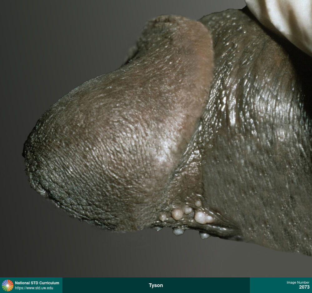
Tyson's glands
Tyson’s glands are white-yellow bumps on the underside of the penis, typically on both sides of the frenulum. Tyson’s glans are benign ectopic sebaceous glands that do not require any treatment.
Photo: Papule / Papules, Dark skin tone, Penis
Courtesy of Sue Szabo, PA-C
Papule / Papules Dark skin tone, Penis
2073
No images match this Condition, Description, and Type
Start by making a choice
Condition
Select a condition to view clinical manifestations associated with a diagnosis or condition
Description
Select one or more of the three categories (location on the body, visible clinical signs, and patient-reported symptoms) and descriptors using the filters
Type
View all results, or filter by clinical photos or illustrations